5F4H | pdb_00005f4h

Archael RuvB-like Holiday junction helicase


Experimental Data Snapshot

  • Method: X-RAY DIFFRACTION
  • Resolution: 2.70 Å
  • R-Value Free: 
    0.265 (Depositor), 0.263 (DCC) 
  • R-Value Work: 
    0.227 (Depositor), 0.227 (DCC) 
  • R-Value Observed: 
    0.229 (Depositor) 

wwPDB Validation 3D Report Full Report

Validation slider image for 5F4H

This is version 1.3 of the entry. See complete history

Literature

Structure and Function of a Novel ATPase that Interacts with Holliday Junction Resolvase Hjc and Promotes Branch Migration.

Zhai, B.DuPrez, K.Doukov, T.I.Li, H.Huang, M.Shang, G.Ni, J.Gu, L.Shen, Y.Fan, L.

(2017) J Mol Biology 429: 1009-1029

  • DOI: https://doi.org/10.1016/j.jmb.2017.02.016
  • Primary Citation Related Structures: 
    5F4H

  • PubMed Abstract: 

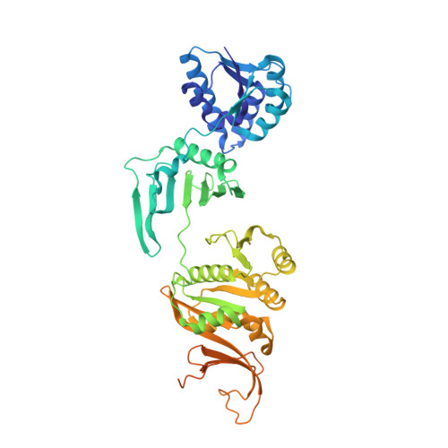
    Holliday junction (HJ) is a hallmark intermediate in DNA recombination and must be processed by dissolution (for double HJ) or resolution to ensure genome stability. Although HJ resolvases have been identified in all domains of life, there is a long-standing effort to search in prokaryotes and eukarya for proteins promoting HJ migration. Here, we report the structural and functional characterization of a novel ATPase, Sulfolobus islandicusPilT N-terminal-domain-containing ATPase (SisPINA), encoded by the gene adjacent to the resolvase Hjc coding gene. PINA is conserved in archaea and vital for S. islandicus viability. Purified SisPINA forms hexameric rings in the crystalline state and in solution, similar to the HJ migration helicase RuvB in Gram-negative bacteria. Structural analysis suggests that ATP binding and hydrolysis cause conformational changes in SisPINA to drive branch migration. Further studies reveal that SisPINA interacts with SisHjc and coordinates HJ migration and cleavage.


  • Organizational Affiliation
    • State Key Laboratory of Microbial Technology, Shandong University, 27 Shanda Nan Road, Jinan 250100, PR China.

Macromolecule Content 

  • Total Structure Weight: 346.03 kDa 
  • Atom Count: 20,358 
  • Modeled Residue Count: 2,616 
  • Deposited Residue Count: 3,030 
  • Unique protein chains: 1

Macromolecules

Find similar proteins by:|  3D Structure
Entity ID: 1
MoleculeChains  Sequence LengthOrganismDetailsImage
Nucleotide binding protein PINc
A, B, C, D, E
A, B, C, D, E, F
505Saccharolobus islandicus L.S.2.15Mutation(s): 1 
Gene Names: LS215_1665
EC: 3.6.4
UniProt
Find proteins for F0NID4 (Saccharolobus islandicus (strain REY15A))
Explore F0NID4 
Go to UniProtKB:  F0NID4
Entity Groups
Sequence Clusters30% Identity50% Identity70% Identity90% Identity95% Identity100% Identity
UniProt GroupF0NID4
Sequence Annotations
Expand
Reference Sequence

Small Molecules

Ligands 2 Unique
IDChains Name / Formula / InChI Key2D Diagram3D Interactions
GOL

Query on GOL


Download:Ideal Coordinates CCD File
BA [auth E]
CA [auth E]
DA [auth E]
EA [auth E]
FA [auth E]
BA [auth E],
CA [auth E],
DA [auth E],
EA [auth E],
FA [auth E],
G [auth A],
GA [auth F],
H [auth A],
HA [auth F],
I [auth A],
IA [auth F],
JA [auth F],
L [auth B],
M [auth B],
R [auth C],
T [auth D],
U [auth D],
V [auth D],
W [auth D],
X [auth D],
Y [auth D]
GLYCEROL
C3 H8 O3
PEDCQBHIVMGVHV-UHFFFAOYSA-N
EDO

Query on EDO


Download:Ideal Coordinates CCD File
AA [auth D]
J [auth A]
K [auth A]
KA [auth F]
LA [auth F]
AA [auth D],
J [auth A],
K [auth A],
KA [auth F],
LA [auth F],
N [auth B],
O [auth B],
P [auth B],
Q [auth B],
S [auth C],
Z [auth D]
1,2-ETHANEDIOL
C2 H6 O2
LYCAIKOWRPUZTN-UHFFFAOYSA-N

Experimental Data & Validation

Experimental Data

  • Method: X-RAY DIFFRACTION
  • Resolution: 2.70 Å
  • R-Value Free:  0.265 (Depositor), 0.263 (DCC) 
  • R-Value Work:  0.227 (Depositor), 0.227 (DCC) 
  • R-Value Observed: 0.229 (Depositor) 
Space Group: P 1 21 1
Unit Cell:
Length ( Å )Angle ( ˚ )
a = 101.72α = 90
b = 148.98β = 104.07
c = 122.13γ = 90
Software Package:
Software NamePurpose
REFMACrefinement
PDB_EXTRACTdata extraction
XDSdata reduction
XDSdata scaling
PHASERphasing
Cootmodel building
Blu-Icedata collection

Structure Validation

View Full Validation Report


Entry History 

Deposition Data

Revision History  (Full details and data files)

  • Version 1.0: 2016-12-21
    Type: Initial release
  • Version 1.1: 2017-09-06
    Changes: Author supporting evidence
  • Version 1.2: 2017-11-01
    Changes: Database references
  • Version 1.3: 2024-10-23
    Changes: Data collection, Database references, Refinement description, Structure summary