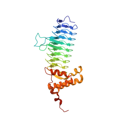
Crystal structure and activity of Francisella novicida UDP-N-acetylglucosamine acyltransferase
Joo, S.H., Chung, H.S.(2016) Biochem Biophys Res Commun 478: 1223-1229
- PubMed: 27545601
- DOI: https://doi.org/10.1016/j.bbrc.2016.08.098
- Primary Citation of Related Structures:
5F42 - PubMed Abstract:
The first step of lipid A biosynthesis in Escherichia coli (E. coli) is catalyzed by LpxA (EcLpxA), an acyltransferase selective for UDP-N-acetylglucosamine (UDP-GlcNAc) and R-3-hydroxymyristoyl-acyl carrier protein (3-OH-C14-ACP), and is an essential step in majority of Gram-negative bacteria. Since the majority of lipid A species isolated from F. novicida contains 3-OH-C16 or 3-OH-C18 at its C3 and C3' positions, FnLpxA was thought to be selective for longer acyl chain (3-OH-C16 and 3-OH-C18) over short acyl chain (3-OH-C14, 3-OH-C12, and 3-OH-C10). Here we demonstrate that Francisella novicida (F. novicida) lpxA functionally complements an E. coli lpxA knockout mutant and efficiently transfers 3-OH-C14 as well as 3-OH-C16 in E. coli. Our results implicate that the acyl chain length of lipid A is determined by several factors including acyl chain selectivity of LpxA and downstream enzymes, as well as the composition of the acyl-ACP pool in vivo. We also report the crystal structure of F. novicida LpxA (FnLpxA) at 2.06 Å. The N-terminal parallel beta-helix (LβH) and C-terminal alpha-helical domain are similar to other reported structures of LpxAs. However, our structure indicates that the supposed ruler residues for hydrocarbon length, 171L in one monomer and 168H in the adjacent monomer in a functional trimer of FnLpxA, are located just 3.8 Å apart that renders not enough space for binding of 3-OH-C12 or longer acyl chains. This implicates that FnLpxA may have an alternative hydrophobic pocket, or the acyl chain may bend while binding to FnLpxA. In addition, the FnLpxA structure suggests a potential inhibitor binding site for development of antibiotics.
- Department of Pharmacy, Catholic University of Daegu, Gyeongbuk 38430, South Korea; Department of Biochemistry, Duke University Medical Center, Durham, NC 27710, USA.
Organizational Affiliation:


