


Crystal Structure of Periplasmic Dipeptide Transport Protein from Yersinia pestis
Kim, Y., Zhou, M., Shatsman, S., Anderson, W.F., Joachimiak, A.To be published.
Experimental Data Snapshot
wwPDB Validation 3D Report Full Report

Entity ID: 1 | |||||
|---|---|---|---|---|---|
| Molecule | Chains | Sequence Length | Organism | Details | Image |
| Periplasmic dipeptide transport protein | 543 | Yersinia pestis Nepal516 | Mutation(s): 0 Gene Names: YPN_3652 | 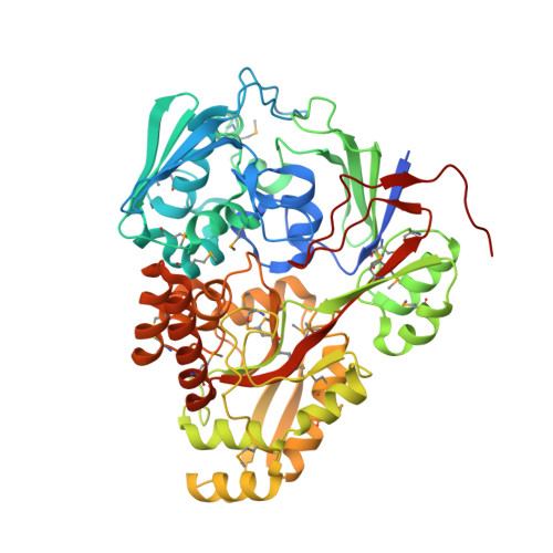 | |
UniProt | |||||
Find proteins for A0A0H2YM47 (Yersinia pestis bv. Antiqua (strain Nepal516)) Explore A0A0H2YM47 Go to UniProtKB: A0A0H2YM47 | |||||
Entity Groups | |||||
| Sequence Clusters | 30% Identity50% Identity70% Identity90% Identity95% Identity100% Identity | ||||
| UniProt Group | A0A0H2YM47 | ||||
Sequence AnnotationsExpand | |||||
Reference Sequence | |||||
| Ligands 5 Unique | |||||
|---|---|---|---|---|---|
| ID | Chains | Name / Formula / InChI Key | 2D Diagram | 3D Interactions | |
| GLN Download:Ideal Coordinates CCD File | J [auth A], K [auth A], R [auth B], S [auth B] | GLUTAMINE C5 H10 N2 O3 ZDXPYRJPNDTMRX-VKHMYHEASA-N |  | ||
| PEG Download:Ideal Coordinates CCD File | F [auth A] | DI(HYDROXYETHYL)ETHER C4 H10 O3 MTHSVFCYNBDYFN-UHFFFAOYSA-N |  | ||
| GOL Download:Ideal Coordinates CCD File | D [auth A] | GLYCEROL C3 H8 O3 PEDCQBHIVMGVHV-UHFFFAOYSA-N |  | ||
| EDO Download:Ideal Coordinates CCD File | C [auth A] E [auth A] G [auth A] I [auth A] L [auth A] | 1,2-ETHANEDIOL C2 H6 O2 LYCAIKOWRPUZTN-UHFFFAOYSA-N |  | ||
| CL Download:Ideal Coordinates CCD File | H [auth A], O [auth B] | CHLORIDE ION Cl VEXZGXHMUGYJMC-UHFFFAOYSA-M |  | ||
| Modified Residues 1 Unique | |||||
|---|---|---|---|---|---|
| ID | Chains | Type | Formula | 2D Diagram | Parent |
| MSE Query on MSE | A, B | L-PEPTIDE LINKING | C5 H11 N O2 Se |  | MET |
| Length ( Å ) | Angle ( ˚ ) |
|---|---|
| a = 289.29 | α = 90 |
| b = 59.745 | β = 103.96 |
| c = 80.631 | γ = 90 |
| Software Name | Purpose |
|---|---|
| PHENIX | refinement |
| HKL-3000 | data scaling |
| HKL-3000 | phasing |
| SBC-Collect | data collection |
| Funding Organization | Location | Grant Number |
|---|---|---|
| National Institutes of Health/National Institute Of Allergy and Infectious Diseases (NIH/NIAID) | United States | -- |


