5EZU | pdb_00005ezu

Crystal structure of the N-terminal domain of vaccinia virus immunomodulator A46 in complex with myristic acid.


Experimental Data Snapshot

  • Method: X-RAY DIFFRACTION
  • Resolution: 1.55 Å
  • R-Value Free: 
    0.242 (Depositor), 0.211 (DCC) 
  • R-Value Work: 
    0.200 (Depositor), 0.201 (DCC) 
  • R-Value Observed: 
    0.202 (Depositor) 

wwPDB Validation 3D Report Full Report

Validation slider image for 5EZU

Ligand Structure Quality Assessment 


This is version 1.3 of the entry. See complete history

Literature

Vaccinia Virus Immunomodulator A46: A Lipid and Protein-Binding Scaffold for Sequestering Host TIR-Domain Proteins.

Fedosyuk, S.Bezerra, G.A.Radakovics, K.Smith, T.K.Sammito, M.Bobik, N.Round, A.Ten Eyck, L.F.Djinovic-Carugo, K.Uson, I.Skern, T.

(2016) PLoS Pathog 12: e1006079-e1006079

  • DOI: https://doi.org/10.1371/journal.ppat.1006079
  • Primary Citation Related Structures: 
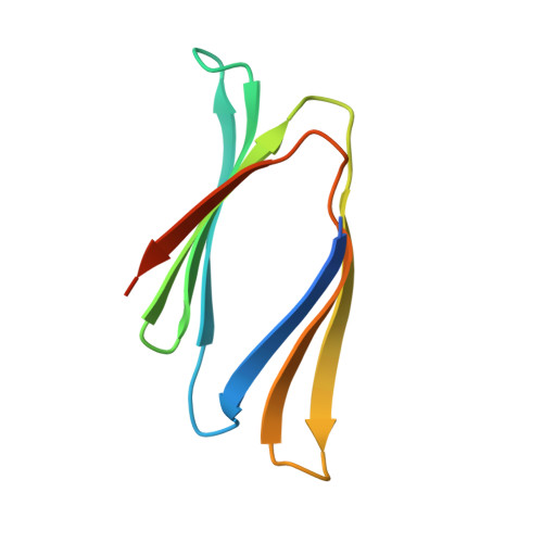
    5EZU

  • PubMed Abstract: 

    Vaccinia virus interferes with early events of the activation pathway of the transcriptional factor NF-kB by binding to numerous host TIR-domain containing adaptor proteins. We have previously determined the X-ray structure of the A46 C-terminal domain; however, the structure and function of the A46 N-terminal domain and its relationship to the C-terminal domain have remained unclear. Here, we biophysically characterize residues 1-83 of the N-terminal domain of A46 and present the X-ray structure at 1.55 Å. Crystallographic phases were obtained by a recently developed ab initio method entitled ARCIMBOLDO_BORGES that employs tertiary structure libraries extracted from the Protein Data Bank; data analysis revealed an all β-sheet structure. This is the first such structure solved by this method which should be applicable to any protein composed entirely of β-sheets. The A46(1-83) structure itself is a β-sandwich containing a co-purified molecule of myristic acid inside a hydrophobic pocket and represents a previously unknown lipid-binding fold. Mass spectrometry analysis confirmed the presence of long-chain fatty acids in both N-terminal and full-length A46; mutation of the hydrophobic pocket reduced the lipid content. Using a combination of high resolution X-ray structures of the N- and C-terminal domains and SAXS analysis of full-length protein A46(1-240), we present here a structural model of A46 in a tetrameric assembly. Integrating affinity measurements and structural data, we propose how A46 simultaneously interferes with several TIR-domain containing proteins to inhibit NF-κB activation and postulate that A46 employs a bipartite binding arrangement to sequester the host immune adaptors TRAM and MyD88.


  • Organizational Affiliation
    • Max F. Perutz Laboratories, Medical University of Vienna, Vienna Biocenter, Dr. Bohr-Gasse 9/3, Vienna, Austria.

Macromolecule Content 

  • Total Structure Weight: 20.3 kDa 
  • Atom Count: 1,276 
  • Modeled Residue Count: 143 
  • Deposited Residue Count: 178 
  • Unique protein chains: 1

Macromolecules

Find similar proteins by:|  3D Structure
Entity ID: 1
MoleculeChains  Sequence LengthOrganismDetailsImage
Protein A46
A, B
89Vaccinia virus WRMutation(s): 0 
Gene Names: VACWR172A46R
UniProt
Find proteins for P26672 (Vaccinia virus (strain Western Reserve))
Explore P26672 
Go to UniProtKB:  P26672
Entity Groups
Sequence Clusters30% Identity50% Identity70% Identity90% Identity95% Identity100% Identity
UniProt GroupP26672
Sequence Annotations
Expand
Reference Sequence

Small Molecules

Ligands 1 Unique
IDChains Name / Formula / InChI Key2D Diagram3D Interactions
MYR

Query on MYR


Download:Ideal Coordinates CCD File
C [auth A]MYRISTIC ACID
C14 H28 O2
TUNFSRHWOTWDNC-UHFFFAOYSA-N

Experimental Data & Validation

Experimental Data

  • Method: X-RAY DIFFRACTION
  • Resolution: 1.55 Å
  • R-Value Free:  0.242 (Depositor), 0.211 (DCC) 
  • R-Value Work:  0.200 (Depositor), 0.201 (DCC) 
  • R-Value Observed: 0.202 (Depositor) 
Space Group: C 1 2 1
Unit Cell:
Length ( Å )Angle ( ˚ )
a = 65.787α = 90
b = 59.58β = 117.7
c = 47.257γ = 90
Software Package:
Software NamePurpose
PHENIXrefinement
PDB_EXTRACTdata extraction
XPREPdata reduction
Arcimboldophasing

Structure Validation

View Full Validation Report


Ligand Structure Quality Assessment 


Entry History 

& Funding Information

Deposition Data


Funding OrganizationLocationGrant Number
Austrian Science FundAustriaW1221

Revision History  (Full details and data files)

  • Version 1.0: 2016-11-30
    Type: Initial release
  • Version 1.1: 2016-12-28
    Changes: Database references
  • Version 1.2: 2017-09-06
    Changes: Author supporting evidence, Refinement description
  • Version 1.3: 2024-05-08
    Changes: Data collection, Database references