5EW7 | pdb_00005ew7

Crystal structure of C9ORF72 Antisense CCCCGG repeat RNA associated with Lou Gehrig's disease and frontotemporal dementia, crystallized with Ba2+


Experimental Data Snapshot

  • Method: X-RAY DIFFRACTION
  • Resolution: 1.75 Å
  • R-Value Free: 
    0.258 (Depositor), 0.258 (DCC) 
  • R-Value Work: 
    0.235 (Depositor), 0.237 (DCC) 
  • R-Value Observed: 
    0.237 (Depositor) 

Starting Model: experimental
View more details

wwPDB Validation 3D Report Full Report

Validation slider image for 5EW7

This is version 1.2 of the entry. See complete history

Literature

Pathogenic C9ORF72 Antisense Repeat RNA Forms a Double Helix with Tandem C:C Mismatches.

Dodd, D.W.Tomchick, D.R.Corey, D.R.Gagnon, K.T.

(2016) Biochemistry 55: 1283-1286

  • DOI: https://doi.org/10.1021/acs.biochem.6b00136
  • Primary Citation Related Structures: 
    5EW4, 5EW7

  • PubMed Abstract: 

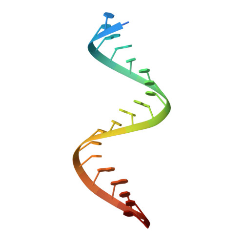
    Expansion of a GGGGCC/CCCCGG repeat sequence in the first intron of the C9ORF72 gene is a leading cause of frontotemporal dementia (FTD) and amyotrophic lateral sclerosis (ALS). In this combined disorder, called c9FTD/ALS, the expansion is bidirectionally transcribed into sense and antisense repeat RNA associated with disease. To better understand the role of C9ORF72 repeat RNA in molecular disease pathology, we determined crystal structures of a [(CCCCGG)3(CCCC)] model antisense repeat RNA to 1.47 Å resolution. The RNA structure was an A-form-like double helix composed of repeating and regularly spaced tandem C:C mismatch pairs that perturbed helical geometry and surface charge. Solution studies revealed a preference for A-form-like helical conformations as the repeat number increased. Results provide a structural starting point for rationalizing the contribution of repeat RNA to c9FTD/ALS molecular disease mechanisms and for developing molecules to target C9ORF72 repeat RNA as potential therapeutics.


  • Organizational Affiliation
    • Department of Pharmacology, ‡Department of Biophysics, and §Department of Biochemistry, University of Texas Southwestern Medical Center , Dallas, Texas 75390, United States.

Macromolecule Content 

  • Total Structure Weight: 14.21 kDa 
  • Atom Count: 927 
  • Modeled Residue Count: 41 
  • Deposited Residue Count: 44 
  • Unique nucleic acid chains: 1

Macromolecules

Find similar nucleic acids by:  Sequence
Entity ID: 1
MoleculeChains LengthOrganismImage
RNA (5'-R(*CP*CP*CP*CP*GP*GP*CP*CP*CP*CP*GP*GP*CP*CP*CP*CP*GP*GP*CP*CP*CP*C)-3')
A, B
22Homo sapiens
Sequence Annotations
Expand
Reference Sequence

Small Molecules

Ligands 3 Unique
IDChains Name / Formula / InChI Key2D Diagram3D Interactions
BA

Query on BA


Download:Ideal Coordinates CCD File
G [auth B]BARIUM ION
Ba
XDFCIPNJCBUZJN-UHFFFAOYSA-N
K

Query on K


Download:Ideal Coordinates CCD File
C [auth A],
H [auth B],
I [auth B],
J [auth B]
POTASSIUM ION
K
NPYPAHLBTDXSSS-UHFFFAOYSA-N
MG

Query on MG


Download:Ideal Coordinates CCD File
D [auth A],
E [auth A],
F [auth A],
K [auth B]
MAGNESIUM ION
Mg
JLVVSXFLKOJNIY-UHFFFAOYSA-N

Experimental Data & Validation

Experimental Data

  • Method: X-RAY DIFFRACTION
  • Resolution: 1.75 Å
  • R-Value Free:  0.258 (Depositor), 0.258 (DCC) 
  • R-Value Work:  0.235 (Depositor), 0.237 (DCC) 
  • R-Value Observed: 0.237 (Depositor) 
Space Group: P 21 21 21
Unit Cell:
Length ( Å )Angle ( ˚ )
a = 41.134α = 90
b = 47.659β = 90
c = 59.495γ = 90
Software Package:
Software NamePurpose
PHENIXrefinement
HKL-3000data scaling
PDB_EXTRACTdata extraction
HKL-3000data reduction
PHENIXphasing

Structure Validation

View Full Validation Report


Entry History 

Deposition Data

Revision History  (Full details and data files)

  • Version 1.0: 2016-02-24
    Type: Initial release
  • Version 1.1: 2016-03-16
    Changes: Database references
  • Version 1.2: 2023-09-27
    Changes: Advisory, Data collection, Database references, Derived calculations, Refinement description