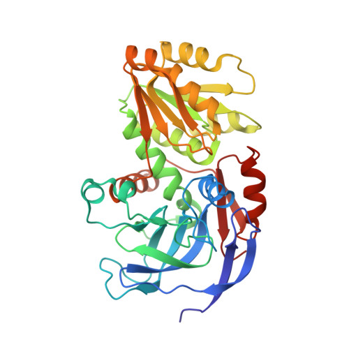
5ENV

YEAST ALCOHOL DEHYDROGENASE WITH BOUND COENZYME


Experimental Data Snapshot

  • Method: X-RAY DIFFRACTION
  • Resolution: 3.00 Å
  • R-Value Free: 0.197 
  • R-Value Work: 0.132 
  • R-Value Observed: 0.135 

Starting Model: experimental
View more details

wwPDB Validation   3D Report Full Report


Ligand Structure Quality Assessment 


This is version 1.5 of the entry. See complete history


Literature

Mechanistic implications from structures of yeast alcohol dehydrogenase complexed with coenzyme and an alcohol.

Plapp, B.V.Charlier, H.A.Ramaswamy, S.

(2015) Arch Biochem Biophys 591: 35-42

  • DOI: https://doi.org/10.1016/j.abb.2015.12.009
  • Primary Citation of Related Structures:  
    5ENV

  • PubMed Abstract: 

    Yeast alcohol dehydrogenase I is a homotetramer of subunits with 347 amino acid residues, catalyzing the oxidation of alcohols using NAD(+) as coenzyme. A new X-ray structure was determined at 3.0 Å where both subunits of an asymmetric dimer bind coenzyme and trifluoroethanol. The tetramer is a pair of back-to-back dimers. Subunit A has a closed conformation and can represent a Michaelis complex with an appropriate geometry for hydride transfer between coenzyme and alcohol, with the oxygen of 2,2,2-trifluoroethanol ligated at 2.1 Å to the catalytic zinc in the classical tetrahedral coordination with Cys-43, Cys-153, and His-66. Subunit B has an open conformation, and the coenzyme interacts with amino acid residues from the coenzyme binding domain, but not with residues from the catalytic domain. Coenzyme appears to bind to and dissociate from the open conformation. The catalytic zinc in subunit B has an alternative, inverted coordination with Cys-43, Cys-153, His-66 and the carboxylate of Glu-67, while the oxygen of trifluoroethanol is 3.5 Å from the zinc. Subunit B may represent an intermediate in the mechanism after coenzyme and alcohol bind and before the conformation changes to the closed form and the alcohol oxygen binds to the zinc and displaces Glu-67.


  • Organizational Affiliation

    Department of Biochemistry, The University of Iowa, Iowa City, IA 52242, USA. Electronic address: bv-plapp@uiowa.edu.


Macromolecules
Find similar proteins by:  (by identity cutoff)  |  3D Structure
Entity ID: 1
MoleculeChains Sequence LengthOrganismDetailsImage
Alcohol dehydrogenase 1
A, B
347Saccharomyces cerevisiae S288CMutation(s): 0 
Gene Names: ADH1ADC1YOL086CO0947
EC: 1.1.1.1 (PDB Primary Data), 1.1.1.78 (UniProt), 1.1.1.54 (UniProt)
UniProt
Find proteins for P00330 (Saccharomyces cerevisiae (strain ATCC 204508 / S288c))
Explore P00330 
Go to UniProtKB:  P00330
Entity Groups  
Sequence Clusters30% Identity50% Identity70% Identity90% Identity95% Identity100% Identity
UniProt GroupP00330
Sequence Annotations
Expand
  • Reference Sequence
Small Molecules
Ligands 3 Unique
IDChains Name / Formula / InChI Key2D Diagram3D Interactions
NAD
Query on NAD

Download Ideal Coordinates CCD File 
E [auth A],
I [auth B]
NICOTINAMIDE-ADENINE-DINUCLEOTIDE
C21 H27 N7 O14 P2
BAWFJGJZGIEFAR-NNYOXOHSSA-N
ETF
Query on ETF

Download Ideal Coordinates CCD File 
F [auth A],
J [auth B]
TRIFLUOROETHANOL
C2 H3 F3 O
RHQDFWAXVIIEBN-UHFFFAOYSA-N
ZN
Query on ZN

Download Ideal Coordinates CCD File 
C [auth A],
D [auth A],
G [auth B],
H [auth B]
ZINC ION
Zn
PTFCDOFLOPIGGS-UHFFFAOYSA-N
Experimental Data & Validation

Experimental Data

  • Method: X-RAY DIFFRACTION
  • Resolution: 3.00 Å
  • R-Value Free: 0.197 
  • R-Value Work: 0.132 
  • R-Value Observed: 0.135 
  • Space Group: P 3 2 1
Unit Cell:
Length ( Å )Angle ( ˚ )
a = 146.256α = 90
b = 146.256β = 90
c = 65.98γ = 120
Software Package:
Software NamePurpose
REFMACrefinement
DENZOdata reduction
SCALEPACKdata scaling
MOLREPphasing

Structure Validation

View Full Validation Report


Ligand Structure Quality Assessment 


Entry History & Funding Information

Deposition Data


Funding OrganizationLocationGrant Number
National Science Foundation (NSF, United States)United StatesMCB 95-06831

Revision History  (Full details and data files)

  • Version 1.0: 2015-11-25
    Type: Initial release
  • Version 1.1: 2016-01-20
    Changes: Database references
  • Version 1.2: 2017-09-27
    Changes: Author supporting evidence, Data collection, Database references, Derived calculations
  • Version 1.3: 2019-11-27
    Changes: Author supporting evidence, Data collection
  • Version 1.4: 2023-09-27
    Changes: Data collection, Database references, Derived calculations, Refinement description
  • Version 1.5: 2024-10-16
    Changes: Structure summary