5EN9

High resolution x-ray crystal structure of isotope-labeled ester-insulin


Experimental Data Snapshot

  • Method: X-RAY DIFFRACTION
  • Resolution: 1.50 Å
  • R-Value Free: 0.177 
  • R-Value Work: 0.143 
  • R-Value Observed: 0.146 

wwPDB Validation   3D Report Full Report


This is version 1.4 of the entry. See complete history


Literature

Crystallization of Enantiomerically Pure Proteins from Quasi-Racemic Mixtures: Structure Determination by X-Ray Diffraction of Isotope-Labeled Ester Insulin and Human Insulin.

Mandal, K.Dhayalan, B.Avital-Shmilovici, M.Tokmakoff, A.Kent, S.B.

(2016) Chembiochem 17: 421-425

  • DOI: https://doi.org/10.1002/cbic.201500600
  • Primary Citation of Related Structures:  
    5EN9, 5ENA

  • PubMed Abstract: 

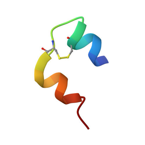
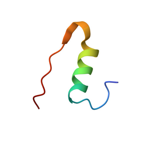
    As a part of a program aimed towards the study of the dynamics of human insulin-protein dimer formation using two-dimensional infrared spectroscopy, we used total chemical synthesis to prepare stable isotope labeled [(1-(13) C=(18) O)Phe(B24) )] human insulin, via [(1-(13) C=(18) O)Phe(B24) )] ester insulin as a key intermediate product that facilitates folding of the synthetic protein molecule (see preceding article). Here, we describe the crystal structure of the synthetic isotope-labeled ester insulin intermediate and the product synthetic human insulin. Additionally, we present our observations on hexamer formation with these two proteins in the absence of phenol derivatives and/or Zn metal ions. We also describe and discuss the fractional crystallization of quasi-racemic protein mixtures containing each of these two synthetic proteins.


  • Organizational Affiliation

    Department of Chemistry, University of Chicago, 929 East 57th Street, Chicago, IL, 60637, USA. kmandalt@uchicago.edu.


Macromolecules

Find similar proteins by:  Sequence   |   3D Structure  

Entity ID: 1
MoleculeChains Sequence LengthOrganismDetailsImage
Insulin chain A21Homo sapiensMutation(s): 0 
UniProt & NIH Common Fund Data Resources
Find proteins for P01308 (Homo sapiens)
Explore P01308 
Go to UniProtKB:  P01308
PHAROS:  P01308
GTEx:  ENSG00000254647 
Entity Groups  
Sequence Clusters30% Identity50% Identity70% Identity90% Identity95% Identity100% Identity
UniProt GroupP01308
Sequence Annotations
Expand
  • Reference Sequence
Find similar proteins by:  (by identity cutoff)  |  3D Structure
Entity ID: 2
MoleculeChains Sequence LengthOrganismDetailsImage
Insulin chain B30Homo sapiensMutation(s): 0 
UniProt & NIH Common Fund Data Resources
Find proteins for P01308 (Homo sapiens)
Explore P01308 
Go to UniProtKB:  P01308
PHAROS:  P01308
GTEx:  ENSG00000254647 
Entity Groups  
Sequence Clusters30% Identity50% Identity70% Identity90% Identity95% Identity100% Identity
UniProt GroupP01308
Sequence Annotations
Expand
  • Reference Sequence
Experimental Data & Validation

Experimental Data

  • Method: X-RAY DIFFRACTION
  • Resolution: 1.50 Å
  • R-Value Free: 0.177 
  • R-Value Work: 0.143 
  • R-Value Observed: 0.146 
  • Space Group: I 21 3
Unit Cell:
Length ( Å )Angle ( ˚ )
a = 77.981α = 90
b = 77.981β = 90
c = 77.981γ = 90
Software Package:
Software NamePurpose
HKL-2000data collection
SCALEPACKdata scaling
PHASERphasing
PHENIXrefinement
PDB_EXTRACTdata extraction

Structure Validation

View Full Validation Report


Entry History & Funding Information

Deposition Data


Funding OrganizationLocationGrant Number
National Institutes of Health/National Institute of Diabetes and Digestive and Kidney Disease (NIH/NIDDK)United StatesR01DK08993

Revision History  (Full details and data files)

  • Version 1.0: 2016-01-13
    Type: Initial release
  • Version 1.1: 2016-01-20
    Changes: Data collection
  • Version 1.2: 2016-03-16
    Changes: Database references
  • Version 1.3: 2017-09-20
    Changes: Author supporting evidence, Derived calculations
  • Version 1.4: 2019-12-25
    Changes: Author supporting evidence