


Binding of Mg2+ATP Enhances Inhibition of Human Mitochondrial Tryptophanyl-tRNA Synthetase by Indolmycin
Williams, T.L., Carter Jr., C.W.To be published.
Experimental Data Snapshot
Starting Model: experimental
View more details
Entity ID: 1 | |||||
|---|---|---|---|---|---|
| Molecule | Chains | Sequence Length | Organism | Details | Image |
| Tryptophan--tRNA ligase, mitochondrial | 343 | Homo sapiens | Mutation(s): 0 Gene Names: WARS2 EC: 6.1.1.2 | 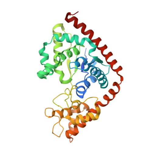 | |
UniProt & NIH Common Fund Data Resources | |||||
PHAROS: Q9UGM6 GTEx: ENSG00000116874 | |||||
Entity Groups | |||||
| Sequence Clusters | 30% Identity50% Identity70% Identity90% Identity95% Identity100% Identity | ||||
| UniProt Group | Q9UGM6 | ||||
Sequence AnnotationsExpand | |||||
Reference Sequence | |||||
| Ligands 4 Unique | |||||
|---|---|---|---|---|---|
| ID | Chains | Name / Formula / InChI Key | 2D Diagram | 3D Interactions | |
| ATP Download:Ideal Coordinates CCD File | D [auth A], H [auth B] | ADENOSINE-5'-TRIPHOSPHATE C10 H16 N5 O13 P3 ZKHQWZAMYRWXGA-KQYNXXCUSA-N |  | ||
| 5BX Download:Ideal Coordinates CCD File | C [auth A], G [auth B] | (5S)-5-[(1R)-1-(1H-indol-3-yl)ethyl]-2-(methylamino)-1,3-oxazol-4(5H)-one C14 H15 N3 O2 GNTVWGDQPXCYBV-PELKAZGASA-N |  | ||
| GOL Download:Ideal Coordinates CCD File | K [auth B], L [auth B] | GLYCEROL C3 H8 O3 PEDCQBHIVMGVHV-UHFFFAOYSA-N |  | ||
| MN Download:Ideal Coordinates CCD File | E [auth A], F [auth A], I [auth B], J [auth B] | MANGANESE (II) ION Mn WAEMQWOKJMHJLA-UHFFFAOYSA-N |  | ||
| Length ( Å ) | Angle ( ˚ ) |
|---|---|
| a = 58.289 | α = 90 |
| b = 78.087 | β = 96.55 |
| c = 153.996 | γ = 90 |
| Software Name | Purpose |
|---|---|
| PHENIX | refinement |
| SCALA | data scaling |
| Coot | model building |
| Funding Organization | Location | Grant Number |
|---|---|---|
| National Institutes of Health/National Institute of General Medical Sciences (NIH/NIGMS) | United States | GM40906 |


