


The structural and biochemical characterization of acyl-coa hydrolase from Staphylococcus aureus
Khandokar, Y.B., Srivastava, P.S., Forwood, J.K.To be published.
Experimental Data Snapshot
Starting Model: experimental
View more details
Entity ID: 1 | |||||
|---|---|---|---|---|---|
| Molecule | Chains | Sequence Length | Organism | Details | Image |
| Acyl CoA Hydrolase | 176 | Staphylococcus aureus subsp. aureus Mu50 | Mutation(s): 0 Gene Names: SAV1878 | 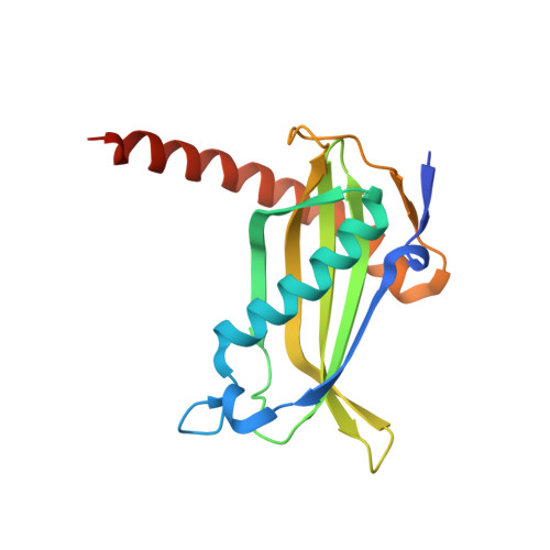 | |
UniProt | |||||
Find proteins for A0A0H3K033 (Staphylococcus aureus (strain Mu50 / ATCC 700699)) Explore A0A0H3K033 Go to UniProtKB: A0A0H3K033 | |||||
Entity Groups | |||||
| Sequence Clusters | 30% Identity50% Identity70% Identity90% Identity95% Identity100% Identity | ||||
| UniProt Group | A0A0H3K033 | ||||
Sequence AnnotationsExpand | |||||
Reference Sequence | |||||
| Ligands 3 Unique | |||||
|---|---|---|---|---|---|
| ID | Chains | Name / Formula / InChI Key | 2D Diagram | 3D Interactions | |
| 5NG Download:Ideal Coordinates CCD File | F [auth C] | [[(2~{S},3~{S},4~{R},5~{R})-5-(6-aminopurin-9-yl)-4-oxidanyl-3-phosphonooxy-oxolan-2-yl]methoxy-oxidanyl-phosphoryl] [(3~{R})-4-[[3-[2-[2-[3-[[(2~{R})-4-[[[(2~{R},3~{S},4~{R},5~{R})-5-(6-aminopurin-9-yl)-4-oxidanyl-3-phosphonooxy-oxolan-2-yl]methoxy-oxidanyl-phosphoryl]oxy-oxidanyl-phosphoryl]oxy-3,3-dimethyl-2-oxidanyl-butanoyl]amino]propanoylamino]ethyldisulfanyl]ethylamino]-3-oxidanylidene-propyl]amino]-2,2-dimethyl-3-oxidanyl-4-oxidanylidene-butyl] hydrogen phosphate C42 H70 N14 O32 P6 S2 YAISMNQCMHVVLO-BJLFRJKCSA-N |  | ||
| BCO Download:Ideal Coordinates CCD File | D [auth A] | Butyryl Coenzyme A C25 H42 N7 O17 P3 S CRFNGMNYKDXRTN-CITAKDKDSA-N |  | ||
| COA Download:Ideal Coordinates CCD File | E [auth B] | COENZYME A C21 H36 N7 O16 P3 S RGJOEKWQDUBAIZ-IBOSZNHHSA-N |  | ||
| Length ( Å ) | Angle ( ˚ ) |
|---|---|
| a = 142.26 | α = 90 |
| b = 142.26 | β = 90 |
| c = 163.892 | γ = 120 |
| Software Name | Purpose |
|---|---|
| PHENIX | refinement |
| iMOSFLM | data reduction |
| Aimless | data scaling |
| PHASER | phasing |


