5EE3 | pdb_00005ee3

COMPLEX STRUCTURE OF OSYCHF1 WITH AMP-PNP


Experimental Data Snapshot

  • Method: X-RAY DIFFRACTION
  • Resolution: 2.90 Å
  • R-Value Free: 
    0.261 (Depositor), 0.259 (DCC) 
  • R-Value Work: 
    0.227 (Depositor), 0.226 (DCC) 
  • R-Value Observed: 
    0.229 (Depositor) 

wwPDB Validation 3D Report Full Report

Validation slider image for 5EE3

Ligand Structure Quality Assessment 


This is version 1.2 of the entry. See complete history

Literature

ATP binding by the P-loop NTPase OsYchF1 (an unconventional G protein) contributes to biotic but not abiotic stress responses

Cheung, M.-Y.Li, X.Miao, R.Fong, Y.-H.Li, K.-P.Yung, Y.-L.Yu, M.-H.Wong, K.-B.Chen, Z.Lam, H.-M.

(2016) Proc Natl Acad Sci U S A 113: 2648-2653

  • DOI: https://doi.org/10.1073/pnas.1522966113
  • Primary Citation Related Structures: 
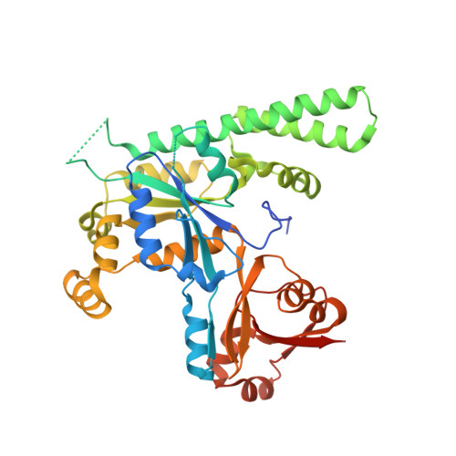
    5EE0, 5EE1, 5EE3, 5EE9

  • PubMed Abstract: 

    G proteins are involved in almost all aspects of the cellular regulatory pathways through their ability to bind and hydrolyze GTP. The YchF subfamily, interestingly, possesses the unique ability to bind both ATP and GTP, and is possibly an ancestral form of G proteins based on phylogenetic studies and is present in all kingdoms of life. However, the biological significance of such a relaxed ligand specificity has long eluded researchers. Here, we have elucidated the different conformational changes caused by the binding of a YchF homolog in rice (OsYchF1) to ATP versus GTP by X-ray crystallography. Furthermore, by comparing the 3D relationships of the ligand position and the various amino acid residues at the binding sites in the crystal structures of the apo-bound and ligand-bound versions, a mechanism for the protein's ability to bind both ligands is revealed. Mutation of the noncanonical G4 motif of the OsYchF1 to the canonical sequence for GTP specificity precludes the binding/hydrolysis of ATP and prevents OsYchF1 from functioning as a negative regulator of plant-defense responses, while retaining its ability to bind/hydrolyze GTP and its function as a negative regulator of abiotic stress responses, demonstrating the specific role of ATP-binding/hydrolysis in disease resistance. This discovery will have a significant impact on our understanding of the structure-function relationships of the YchF subfamily of G proteins in all kingdoms of life.


  • Organizational Affiliation
    • School of Life Sciences, The Chinese University of Hong Kong, Shatin, N.T., Hong Kong SAR; Center for Soybean Research of the State Key Laboratory of Agrobiotechnology, The Chinese University of Hong Kong, Shatin, N.T., Hong Kong SAR;

Macromolecule Content 

  • Total Structure Weight: 90.58 kDa 
  • Atom Count: 5,604 
  • Modeled Residue Count: 721 
  • Deposited Residue Count: 790 
  • Unique protein chains: 1

Macromolecules

Find similar proteins by:|  3D Structure
Entity ID: 1
MoleculeChains  Sequence LengthOrganismDetailsImage
Obg-like ATPase 1
A, B
395Oryza sativa Japonica GroupMutation(s): 0 
Gene Names: OS08G0199300OSYCHF1
EC: 3.6.5
UniProt
Find proteins for Q6Z1J6 (Oryza sativa subsp. japonica)
Explore Q6Z1J6 
Go to UniProtKB:  Q6Z1J6
Entity Groups
Sequence Clusters30% Identity50% Identity70% Identity90% Identity95% Identity100% Identity
UniProt GroupQ6Z1J6
Sequence Annotations
Expand
Reference Sequence

Small Molecules

Ligands 5 Unique
IDChains Name / Formula / InChI Key2D Diagram3D Interactions
ANP

Query on ANP


Download:Ideal Coordinates CCD File
C [auth A],
I [auth B]
PHOSPHOAMINOPHOSPHONIC ACID-ADENYLATE ESTER
C10 H17 N6 O12 P3
PVKSNHVPLWYQGJ-KQYNXXCUSA-N
EPE

Query on EPE


Download:Ideal Coordinates CCD File
H [auth A]4-(2-HYDROXYETHYL)-1-PIPERAZINE ETHANESULFONIC ACID
C8 H18 N2 O4 S
JKMHFZQWWAIEOD-UHFFFAOYSA-N
SO4

Query on SO4


Download:Ideal Coordinates CCD File
N [auth B]SULFATE ION
O4 S
QAOWNCQODCNURD-UHFFFAOYSA-L
CL

Query on CL


Download:Ideal Coordinates CCD File
E [auth A]
F [auth A]
G [auth A]
K [auth B]
L [auth B]
E [auth A],
F [auth A],
G [auth A],
K [auth B],
L [auth B],
M [auth B]
CHLORIDE ION
Cl
VEXZGXHMUGYJMC-UHFFFAOYSA-M
MG

Query on MG


Download:Ideal Coordinates CCD File
D [auth A],
J [auth B]
MAGNESIUM ION
Mg
JLVVSXFLKOJNIY-UHFFFAOYSA-N

Experimental Data & Validation

Experimental Data

  • Method: X-RAY DIFFRACTION
  • Resolution: 2.90 Å
  • R-Value Free:  0.261 (Depositor), 0.259 (DCC) 
  • R-Value Work:  0.227 (Depositor), 0.226 (DCC) 
  • R-Value Observed: 0.229 (Depositor) 
Space Group: P 21 21 21
Unit Cell:
Length ( Å )Angle ( ˚ )
a = 70.086α = 90
b = 112.39β = 90
c = 115.023γ = 90
Software Package:
Software NamePurpose
REFMACrefinement
HKL-2000data reduction
HKL-2000data scaling
BALBESphasing

Structure Validation

View Full Validation Report


Ligand Structure Quality Assessment 


Entry History 

Deposition Data

Revision History  (Full details and data files)

  • Version 1.0: 2016-02-24
    Type: Initial release
  • Version 1.1: 2016-03-23
    Changes: Database references
  • Version 1.2: 2024-03-20
    Changes: Data collection, Database references, Derived calculations