5EDM | pdb_00005edm

Crystal structure of prothrombin deletion mutant residues 154-167 ( Form I )


Experimental Data Snapshot

  • Method: X-RAY DIFFRACTION
  • Resolution: 2.20 Å
  • R-Value Free: 
    0.236 (Depositor), 0.238 (DCC) 
  • R-Value Work: 
    0.196 (Depositor), 0.205 (DCC) 
  • R-Value Observed: 
    0.198 (Depositor) 

Starting Model: experimental
View more details

wwPDB Validation 3D Report Full Report

Validation slider image for 5EDM

Ligand Structure Quality Assessment 


This is version 2.2 of the entry. See complete history

Literature

How the Linker Connecting the Two Kringles Influences Activation and Conformational Plasticity of Prothrombin.

Pozzi, N.Chen, Z.Di Cera, E.

(2016) J Biological Chem 291: 6071-6082

  • DOI: https://doi.org/10.1074/jbc.M115.700401
  • Primary Citation Related Structures: 
    5EDK, 5EDM

  • PubMed Abstract: 

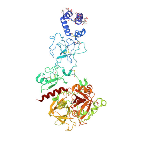
    A flexible linker (Lnk2) composed of 26 amino acids connects kringle-1 to kringle-2 in the coagulation factor prothrombin. Recent studies point to Lnk2 as a key determinant of the structure and function of this zymogen. Using a combination of mutagenesis, structural biology, and single molecule spectroscopy, we show how Lnk2 influences activation and conformational plasticity of prothrombin. Scrambling the sequence of Lnk2 is inconsequential on activation, and so is extension by as many as 22 residues. On the other hand, below a critical length of 15 residues, the rate of prothrombin activation increases (10-fold) in the absence of cofactor Va and decreases (3-fold) in the presence of cofactor. Furthermore, activation by prothrombinase takes place without preference along the prethrombin-2 (cleavage at Arg(271) first) or meizothrombin (cleavage at Arg(320) first) pathways. Notably, these transitions in the rate and pathway of activation require the presence of phospholipids, pointing to an important physiological role for Lnk2 when prothrombin is anchored to the membrane. Two new crystal structures of prothrombin lacking 22 (ProTΔ146-167) or 14 (ProTΔ154-167) residues of Lnk2 document striking conformational rearrangements of domains located across this linker. FRET measurements of freely diffusing single molecules prove that these structural transitions are genuine properties of the zymogen in solution. These findings support a molecular model of prothrombin activation where Lnk2 presents the sites of cleavage at Arg(271) and Arg(320) to factor Xa in different orientations by pivoting the C-terminal kringle-2/protease domain pair on the N-terminal Gla domain/kringle-1 pair anchored to the membrane.


  • Organizational Affiliation
    • From the Edward A. Doisy Department of Biochemistry and Molecular Biology, Saint Louis University School of Medicine, St. Louis, Missouri 63104.

Macromolecule Content 

  • Total Structure Weight: 67.01 kDa 
  • Atom Count: 5,005 
  • Modeled Residue Count: 550 
  • Deposited Residue Count: 568 
  • Unique protein chains: 1

Macromolecules

Find similar proteins by:|  3D Structure
Entity ID: 1
MoleculeChains  Sequence LengthOrganismDetailsImage
Prothrombin568Homo sapiensMutation(s): 0 
Gene Names: F2
EC: 3.4.21.5
Membrane Entity: Yes 
UniProt & NIH Common Fund Data Resources
Find proteins for P00734 (Homo sapiens)
Explore P00734 
Go to UniProtKB:  P00734
PHAROS:  P00734
GTEx:  ENSG00000180210 
Entity Groups
Sequence Clusters30% Identity50% Identity70% Identity90% Identity95% Identity100% Identity
UniProt GroupP00734
Glycosylation
Glycosylation Sites: 3Go to GlyGen: P00734-1
Sequence Annotations
Expand
Reference Sequence

Oligosaccharides

Help  
Entity ID: 2
MoleculeChains Length2D Diagram GlycosylationD Interactions
2-acetamido-2-deoxy-beta-D-glucopyranose-(1-4)-2-acetamido-2-deoxy-beta-D-glucopyranose
B, C
2N-Glycosylation
Glycosylation Resources
GlyTouCan: G42666HT
GlyCosmos: G42666HT
GlyGen: G42666HT

Small Molecules

Ligands 4 Unique
IDChains Name / Formula / InChI Key2D Diagram3D Interactions
NAG

Query on NAG


Download:Ideal Coordinates CCD File
J [auth A]2-acetamido-2-deoxy-beta-D-glucopyranose
C8 H15 N O6
OVRNDRQMDRJTHS-FMDGEEDCSA-N
SO4

Query on SO4


Download:Ideal Coordinates CCD File
K [auth A]
L [auth A]
M [auth A]
N [auth A]
O [auth A]
K [auth A],
L [auth A],
M [auth A],
N [auth A],
O [auth A],
P [auth A],
Q [auth A],
R [auth A],
T [auth A]
SULFATE ION
O4 S
QAOWNCQODCNURD-UHFFFAOYSA-L
GOL

Query on GOL


Download:Ideal Coordinates CCD File
S [auth A]GLYCEROL
C3 H8 O3
PEDCQBHIVMGVHV-UHFFFAOYSA-N
MG

Query on MG


Download:Ideal Coordinates CCD File
D [auth A]
E [auth A]
F [auth A]
G [auth A]
H [auth A]
D [auth A],
E [auth A],
F [auth A],
G [auth A],
H [auth A],
I [auth A]
MAGNESIUM ION
Mg
JLVVSXFLKOJNIY-UHFFFAOYSA-N
Modified Residues  1 Unique
IDChains TypeFormula2D DiagramParent
CGU
Query on CGU
A
L-PEPTIDE LINKINGC6 H9 N O6GLU

Experimental Data & Validation

Experimental Data

  • Method: X-RAY DIFFRACTION
  • Resolution: 2.20 Å
  • R-Value Free:  0.236 (Depositor), 0.238 (DCC) 
  • R-Value Work:  0.196 (Depositor), 0.205 (DCC) 
  • R-Value Observed: 0.198 (Depositor) 
Space Group: C 2 2 21
Unit Cell:
Length ( Å )Angle ( ˚ )
a = 109.884α = 90
b = 168.69β = 90
c = 144.315γ = 90
Software Package:
Software NamePurpose
REFMACrefinement
HKL-2000data reduction
HKL-2000data scaling
PHASERphasing

Structure Validation

View Full Validation Report


Ligand Structure Quality Assessment 


Entry History 

& Funding Information

Deposition Data


Funding OrganizationLocationGrant Number
National Institutes of Health/National Heart, Lung, and Blood Institute (NIH/NHLBI)United StatesHL049413, HL073813 and HL112303 (EDC)

Revision History  (Full details and data files)

  • Version 1.0: 2016-01-20
    Type: Initial release
  • Version 1.1: 2016-01-27
    Changes: Database references
  • Version 1.2: 2016-03-30
    Changes: Database references
  • Version 1.3: 2017-08-09
    Changes: Database references, Derived calculations
  • Version 1.4: 2017-09-06
    Changes: Author supporting evidence
  • Version 1.5: 2019-12-04
    Changes: Author supporting evidence
  • Version 2.0: 2020-07-29
    Type: Remediation
    Reason: Carbohydrate remediation
    Changes: Atomic model, Data collection, Derived calculations, Structure summary
  • Version 2.1: 2023-09-27
    Changes: Data collection, Database references, Refinement description, Structure summary
  • Version 2.2: 2023-11-15
    Changes: Data collection