5ECT

Mycobacterium tuberculosis dUTPase G143STOP mutant


Experimental Data Snapshot

  • Method: X-RAY DIFFRACTION
  • Resolution: 1.30 Å
  • R-Value Free: 0.157 
  • R-Value Work: 0.123 
  • R-Value Observed: 0.124 

wwPDB Validation   3D Report Full Report


Ligand Structure Quality Assessment 


This is version 1.2 of the entry. See complete history


Literature

Structural Characterization of Arginine Fingers: Identification of an Arginine Finger for the Pyrophosphatase dUTPases.

Nagy, G.N.Suardiaz, R.Lopata, A.Ozohanics, O.Vekey, K.Brooks, B.R.Leveles, I.Toth, J.Vertessy, B.G.Rosta, E.

(2016) J Am Chem Soc 138: 15035-15045

  • DOI: https://doi.org/10.1021/jacs.6b09012
  • Primary Citation of Related Structures:  
    5ECT, 5EDD

  • PubMed Abstract: 

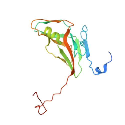
    Arginine finger is a highly conserved and essential residue in many GTPase and AAA+ ATPase enzymes that completes the active site from a distinct protomer, forming contacts with the γ-phosphate of the nucleotide. To date, no pyrophosphatase has been identified that employs an arginine finger fulfilling all of the above properties; all essential arginine fingers are used to catalyze the cleavage of the γ-phosphate. Here, we identify and unveil the role of a conserved arginine residue in trimeric dUTPases that meets all the criteria established for arginine fingers. We found that the conserved arginine adjacent to the P-loop-like motif enables structural organization of the active site for efficient catalysis via its nucleotide coordination, while its direct electrostatic role in transition state stabilization is secondary. An exhaustive structure-based comparison of analogous, conserved arginines from nucleotide hydrolases and transferases revealed a consensus amino acid location and orientation for contacting the γ-phosphate of the substrate nucleotide. Despite the structurally equivalent position, functional differences between arginine fingers of dUTPases and NTPases are explained on the basis of the unique chemistry performed by the pyrophosphatase dUTPases.


  • Organizational Affiliation

    Department of Biotechnology and Food Sciences, Budapest University of Technology and Economics , Budapest 1111, Hungary.


Macromolecules
Find similar proteins by:  (by identity cutoff)  |  3D Structure
Entity ID: 1
MoleculeChains Sequence LengthOrganismDetailsImage
Deoxyuridine 5'-triphosphate nucleotidohydrolase162Mycobacterium tuberculosisMutation(s): 0 
Gene Names: dutRv2697cMTCY05A6.18c
EC: 3.6.1.23
UniProt
Find proteins for P9WNS5 (Mycobacterium tuberculosis (strain ATCC 25618 / H37Rv))
Explore P9WNS5 
Go to UniProtKB:  P9WNS5
Entity Groups  
Sequence Clusters30% Identity50% Identity70% Identity90% Identity95% Identity100% Identity
UniProt GroupP9WNS5
Sequence Annotations
Expand
  • Reference Sequence
Small Molecules
Binding Affinity Annotations 
IDSourceBinding Affinity
DUP Binding MOAD:  5ECT Kd: 5200 (nM) from 1 assay(s)
Experimental Data & Validation

Experimental Data

  • Method: X-RAY DIFFRACTION
  • Resolution: 1.30 Å
  • R-Value Free: 0.157 
  • R-Value Work: 0.123 
  • R-Value Observed: 0.124 
  • Space Group: P 63
Unit Cell:
Length ( Å )Angle ( ˚ )
a = 54.78α = 90
b = 54.78β = 90
c = 84.079γ = 120
Software Package:
Software NamePurpose
REFMACrefinement
XDSdata reduction
XDSdata scaling
Cootmodel building
MOLREPphasing

Structure Validation

View Full Validation Report


Ligand Structure Quality Assessment 


Entry History & Funding Information

Deposition Data


Funding OrganizationLocationGrant Number
Hungarian Scientific Research FundHungaryNK 84008
Hungarian Scientific Research FundHungaryK109486
Biostruct-XHungary283570

Revision History  (Full details and data files)

  • Version 1.0: 2016-11-02
    Type: Initial release
  • Version 1.1: 2016-12-21
    Changes: Database references
  • Version 1.2: 2024-01-10
    Changes: Data collection, Database references, Derived calculations, Refinement description