5ECJ | pdb_00005ecj

Crystal structure of monobody Mb(S4) bound to Prdm14 in complex with Mtgr1


Experimental Data Snapshot

  • Method: X-RAY DIFFRACTION
  • Resolution: 3.05 Å
  • R-Value Free: 
    0.250 (Depositor), 0.254 (DCC) 
  • R-Value Work: 
    0.189 (Depositor), 0.197 (DCC) 
  • R-Value Observed: 
    0.194 (Depositor) 

wwPDB Validation 3D Report Full Report

Validation slider image for 5ECJ

This is version 1.4 of the entry. See complete history

Literature

ETO family protein Mtgr1 mediates Prdm14 functions in stem cell maintenance and primordial germ cell formation.

Nady, N.Gupta, A.Ma, Z.Swigut, T.Koide, A.Koide, S.Wysocka, J.

(2015) Elife 4: e10150-e10150

  • DOI: https://doi.org/10.7554/eLife.10150
  • Primary Citation Related Structures: 
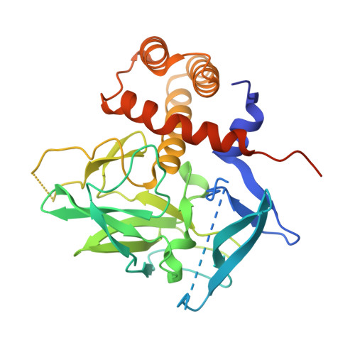
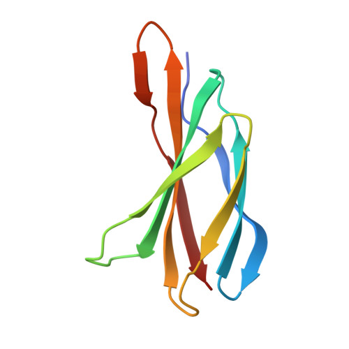
    5ECJ

  • PubMed Abstract: 

    Prdm14 is a sequence-specific transcriptional regulator of embryonic stem cell (ESC) pluripotency and primordial germ cell (PGC) formation. It exerts its function, at least in part, through repressing genes associated with epigenetic modification and cell differentiation. Here, we show that this repressive function is mediated through an ETO-family co-repressor Mtgr1, which tightly binds to the pre-SET/SET domains of Prdm14 and co-occupies its genomic targets in mouse ESCs. We generated two monobodies, synthetic binding proteins, targeting the Prdm14 SET domain and demonstrate their utility, respectively, in facilitating crystallization and structure determination of the Prdm14-Mtgr1 complex, or as genetically encoded inhibitor of the Prdm14-Mtgr1 interaction. Structure-guided point mutants and the monobody abrogated the Prdm14-Mtgr1 association and disrupted Prdm14's function in mESC gene expression and PGC formation in vitro. Altogether, our work uncovers the molecular mechanism underlying Prdm14-mediated repression and provides renewable reagents for studying and controlling Prdm14 functions.


  • Organizational Affiliation
    • Department of Chemical and Systems Biology, Stanford University School of Medicine, Stanford, United States.

Macromolecule Content 

  • Total Structure Weight: 89.44 kDa 
  • Atom Count: 5,480 
  • Modeled Residue Count: 712 
  • Deposited Residue Count: 808 
  • Unique protein chains: 2

Macromolecules

Find similar proteins by:|  3D Structure
Entity ID: 1
MoleculeChains  Sequence LengthOrganismDetailsImage
PR domain zinc finger protein 14,Protein CBFA2T2A,
C [auth B]
309Mus musculusMutation(s): 1 
Gene Names: Prdm14Cbfa2t2Cbfa2t2hMtgr1
EC: 2.1.1
UniProt & NIH Common Fund Data Resources
Find proteins for E9Q3T6 (Mus musculus)
Explore E9Q3T6 
Go to UniProtKB:  E9Q3T6
IMPC:  MGI:3588194
Find proteins for O70374 (Mus musculus)
Explore O70374 
Go to UniProtKB:  O70374
Entity Groups
Sequence Clusters30% Identity50% Identity70% Identity90% Identity95% Identity100% Identity
UniProt GroupsO70374E9Q3T6
Sequence Annotations
Expand
Reference Sequence
Find similar proteins by:|  3D Structure
Entity ID: 2
MoleculeChains  Sequence LengthOrganismDetailsImage
Monobody Mb(S4)B [auth F],
D [auth E]
95Homo sapiensMutation(s): 0 

Experimental Data & Validation

Experimental Data

  • Method: X-RAY DIFFRACTION
  • Resolution: 3.05 Å
  • R-Value Free:  0.250 (Depositor), 0.254 (DCC) 
  • R-Value Work:  0.189 (Depositor), 0.197 (DCC) 
  • R-Value Observed: 0.194 (Depositor) 
Space Group: P 43 21 2
Unit Cell:
Length ( Å )Angle ( ˚ )
a = 106.812α = 90
b = 106.812β = 90
c = 180.83γ = 90
Software Package:
Software NamePurpose
PHENIXrefinement
HKL-3000data reduction
HKL-3000data scaling
PHENIXphasing

Structure Validation

View Full Validation Report


Entry History 

& Funding Information

Deposition Data


Funding OrganizationLocationGrant Number
National Institutes of Health/National Institute on Drug Abuse (NIH/NIDA)United StatesR01DA036887
National Institutes of Health/National Institute of General Medical Sciences (NIH/NIGMS)United StatesR01GM090324

Revision History  (Full details and data files)

  • Version 1.0: 2015-12-02
    Type: Initial release
  • Version 1.1: 2016-05-04
    Changes: Database references
  • Version 1.2: 2017-09-20
    Changes: Author supporting evidence, Derived calculations
  • Version 1.3: 2019-12-11
    Changes: Author supporting evidence
  • Version 1.4: 2024-03-06
    Changes: Data collection, Database references