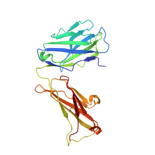
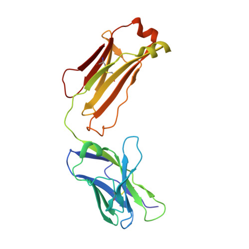
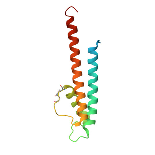
5EC1

KcsA with V76ester mutation


Experimental Data Snapshot

  • Method: X-RAY DIFFRACTION
  • Resolution: 2.75 Å
  • R-Value Free: 0.268 
  • R-Value Work: 0.244 
  • R-Value Observed: 0.246 

Starting Model: experimental
View more details

wwPDB Validation   3D Report Full Report


Ligand Structure Quality Assessment 


This is version 1.5 of the entry. See complete history


Literature

Individual Ion Binding Sites in the K(+) Channel Play Distinct Roles in C-type Inactivation and in Recovery from Inactivation.

Matulef, K.Annen, A.W.Nix, J.C.Valiyaveetil, F.I.

(2016) Structure 24: 750-761

  • DOI: https://doi.org/10.1016/j.str.2016.02.021
  • Primary Citation of Related Structures:  
    5EBL, 5EBM, 5EBW, 5EC1, 5EC2

  • PubMed Abstract: 

    The selectivity filter of K(+) channels contains four ion binding sites (S1-S4) and serves dual functions of discriminating K(+) from Na(+) and acting as a gate during C-type inactivation. C-type inactivation is modulated by ion binding to the selectivity filter sites, but the underlying mechanism is not known. Here we evaluate how the ion binding sites in the selectivity filter of the KcsA channel participate in C-type inactivation and in recovery from inactivation. We use unnatural amide-to-ester substitutions in the protein backbone to manipulate the S1-S3 sites and a side-chain substitution to perturb the S4 site. We develop an improved semisynthetic approach for generating these amide-to-ester substitutions in the selectivity filter. Our combined electrophysiological and X-ray crystallographic analysis of the selectivity filter mutants show that the ion binding sites play specific roles during inactivation and provide insights into the structural changes at the selectivity filter during C-type inactivation.


  • Organizational Affiliation

    Program in Chemical Biology, Department of Physiology and Pharmacology, Oregon Health & Science University, 3181 Southwest Sam Jackson Park Road, Portland, OR 97239, USA.


Macromolecules
Find similar proteins by:  (by identity cutoff)  |  3D Structure
Entity ID: 1
MoleculeChains Sequence LengthOrganismDetailsImage
Antibody Fab Fragment Light Chain219Mus musculusMutation(s): 0 
Entity Groups  
Sequence Clusters30% Identity50% Identity70% Identity90% Identity95% Identity100% Identity
Sequence Annotations
Expand
  • Reference Sequence
Find similar proteins by:  (by identity cutoff)  |  3D Structure
Entity ID: 2
MoleculeChains Sequence LengthOrganismDetailsImage
Antibody Fab Fragment Light Chain209Mus musculusMutation(s): 0 
Entity Groups  
Sequence Clusters30% Identity50% Identity70% Identity90% Identity95% Identity100% Identity
Sequence Annotations
Expand
  • Reference Sequence
Find similar proteins by:  (by identity cutoff)  |  3D Structure
Entity ID: 3
MoleculeChains Sequence LengthOrganismDetailsImage
pH-gated potassium channel KcsA125Streptomyces lividansMutation(s): 5 
Gene Names: kcsAskc1
Membrane Entity: Yes 
UniProt
Find proteins for P0A334 (Streptomyces lividans)
Explore P0A334 
Go to UniProtKB:  P0A334
Entity Groups  
Sequence Clusters30% Identity50% Identity70% Identity90% Identity95% Identity100% Identity
UniProt GroupP0A334
Sequence Annotations
Expand
  • Reference Sequence
Small Molecules
Ligands 3 Unique
IDChains Name / Formula / InChI Key2D Diagram3D Interactions
DGA
Query on DGA

Download Ideal Coordinates CCD File 
E [auth C]DIACYL GLYCEROL
C39 H76 O5
UHUSDOQQWJGJQS-QNGWXLTQSA-N
F09
Query on F09

Download Ideal Coordinates CCD File 
D [auth C]NONAN-1-OL
C9 H20 O
ZWRUINPWMLAQRD-UHFFFAOYSA-N
K
Query on K

Download Ideal Coordinates CCD File 
F [auth C]
G [auth C]
H [auth C]
I [auth C]
J [auth C]
F [auth C],
G [auth C],
H [auth C],
I [auth C],
J [auth C],
K [auth C]
POTASSIUM ION
K
NPYPAHLBTDXSSS-UHFFFAOYSA-N
Experimental Data & Validation

Experimental Data

  • Method: X-RAY DIFFRACTION
  • Resolution: 2.75 Å
  • R-Value Free: 0.268 
  • R-Value Work: 0.244 
  • R-Value Observed: 0.246 
  • Space Group: I 4
Unit Cell:
Length ( Å )Angle ( ˚ )
a = 155.6α = 90
b = 155.6β = 90
c = 75.64γ = 90
Software Package:
Software NamePurpose
XDSdata scaling
PHENIXrefinement
PDB_EXTRACTdata extraction
XDSdata reduction
PHASERphasing

Structure Validation

View Full Validation Report


Ligand Structure Quality Assessment 


Entry History & Funding Information

Deposition Data


Funding OrganizationLocationGrant Number
National Institutes of Health/National Institute of General Medical Sciences (NIH/NIGMS)United States--

Revision History  (Full details and data files)

  • Version 1.0: 2016-04-20
    Type: Initial release
  • Version 1.1: 2016-05-18
    Changes: Database references
  • Version 1.2: 2017-09-27
    Changes: Author supporting evidence, Database references, Derived calculations
  • Version 1.3: 2019-12-04
    Changes: Author supporting evidence
  • Version 1.4: 2023-09-27
    Changes: Data collection, Database references, Derived calculations, Refinement description
  • Version 1.5: 2023-11-15
    Changes: Data collection