5EBZ | pdb_00005ebz

Crystal structure of human IKK1


Experimental Data Snapshot

  • Method: X-RAY DIFFRACTION
  • Resolution: 4.50 Å
  • R-Value Free: 
    0.276 (Depositor), 0.280 (DCC) 
  • R-Value Work: 
    0.238 (Depositor), 0.241 (DCC) 
  • R-Value Observed: 
    0.238 (Depositor) 

wwPDB Validation 3D Report Full Report

Validation slider image for 5EBZ

Ligand Structure Quality Assessment 


This is version 2.1 of the entry. See complete history

Literature

Structural Basis for the Activation of IKK1/ alpha.

Polley, S.Passos, D.O.Huang, D.B.Mulero, M.C.Mazumder, A.Biswas, T.Verma, I.M.Lyumkis, D.Ghosh, G.

(2016) Cell Rep 17: 1907-1914

  • DOI: https://doi.org/10.1016/j.celrep.2016.10.067
  • Primary Citation Related Structures: 
    5EBZ, 5TQW, 5TQX, 5TQY

  • PubMed Abstract: 

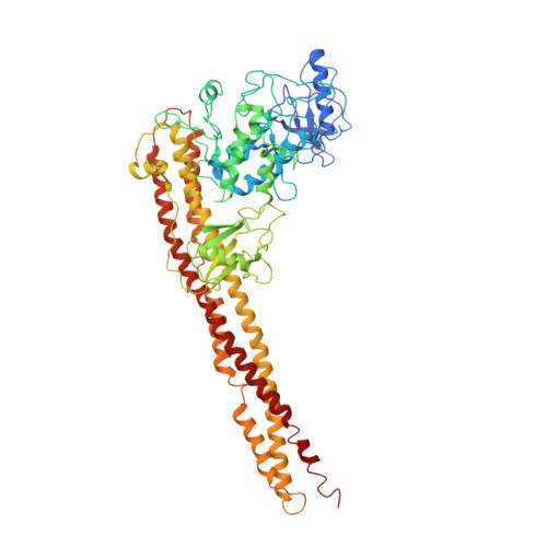
    Distinct signaling pathways activate the NF-κB family of transcription factors. The canonical NF-κB-signaling pathway is mediated by IκB kinase 2/β (IKK2/β), while the non-canonical pathway depends on IKK1/α. The structural and biochemical bases for distinct signaling by these otherwise highly similar IKKs are unclear. We report single-particle cryoelectron microscopy (cryo-EM) and X-ray crystal structures of human IKK1 in dimeric (∼150 kDa) and hexameric (∼450 kDa) forms. The hexamer, which is the representative form in the crystal but comprises only ∼2% of the particles in solution by cryo-EM, is a trimer of IKK1 dimers. While IKK1 hexamers are not detectable in cells, the surface that supports hexamer formation is critical for IKK1-dependent cellular processing of p100 to p52, the hallmark of non-canonical NF-κB signaling. Comparison of this surface to that in IKK2 indicates significant divergence, and it suggests a fundamental role for this surface in signaling by these kinases through distinct pathways.


  • Organizational Affiliation
    • Department of Chemistry & Biochemistry, University of California San Diego, La Jolla, CA 92093, USA; Laboratory of Genetics and Helmsley Center for Genomic Medicine, The Salk Institute for Biological Studies, La Jolla, CA 92037, USA.

Macromolecule Content 

  • Total Structure Weight: 933.78 kDa 
  • Atom Count: 65,132 
  • Modeled Residue Count: 7,860 
  • Deposited Residue Count: 7,860 
  • Unique protein chains: 1

Macromolecules

Find similar proteins by:|  3D Structure
Entity ID: 1
MoleculeChains  Sequence LengthOrganismDetailsImage
Inhibitor of nuclear factor kappa-B kinase subunit alpha
A, B, C, D, E
A, B, C, D, E, F, G, H, I, J, K, L
655Homo sapiensMutation(s): 3 
Gene Names: CHUKIKKATCF16
EC: 2.7.11.10
UniProt & NIH Common Fund Data Resources
Find proteins for O15111 (Homo sapiens)
Explore O15111 
Go to UniProtKB:  O15111
PHAROS:  O15111
GTEx:  ENSG00000213341 
Entity Groups
Sequence Clusters30% Identity50% Identity70% Identity90% Identity95% Identity100% Identity
UniProt GroupO15111
Sequence Annotations
Expand
Reference Sequence

Oligosaccharides

Help  
Entity ID: 3
MoleculeChains Length2D Diagram GlycosylationD Interactions
[(2S,3R,4S,5S,6R)-6-(hydroxymethyl)-2,4-bis(oxidanyl)-5-oxidanylsulfanyloxy-oxan-3-yl] hydrogen sulfate-(1-6)-(2S,3R,4R,5S,6R)-6-(hydroxymethyl)-5-oxidanylsulfanyloxy-oxane-2,3,4-triol-(1-6)-2-O-sulfo-alpha-D-glucopyranose-(1-6)-[(2R,3R,4R,5R,6S)-2-(hydroxymethyl)-5,6-bis(oxidanyl)-3-oxidanylsulfanyloxy-oxan-4-yl] hydrogen sulfate-(1-6)-[(2S,3R,4S,5R,6R)-6-(hydroxymethyl)-2,5-bis(oxidanyl)-4-oxidanylsulfanyloxy-oxan-3-yl] hydrogen sulfate-(1-6)-[(2R,3R,4R,5R,6S)-2-(hydroxymethyl)-5,6-bis(oxidanyl)-3-oxidanylsulfanyloxy-oxan-4-yl] hydrogen sulfate-(1-6)-2,4-di-O-sulfo-alpha-D-glucopyranoseCA [auth c],
IA [auth i],
O,
T
7N/AN/A
Glycosylation Resources
GlyTouCan: G51196UN
GlyCosmos: G51196UN

Small Molecules

Ligands 1 Unique
IDChains Name / Formula / InChI Key2D Diagram3D Interactions
5TL

Query on 5TL


Download:Ideal Coordinates CCD File
OA [auth A]
PA [auth B]
QA [auth C]
RA [auth D]
SA [auth E]
OA [auth A],
PA [auth B],
QA [auth C],
RA [auth D],
SA [auth E],
TA [auth F],
UA [auth G],
VA [auth H],
WA [auth I],
XA [auth J],
YA [auth K],
ZA [auth L]
2-azanyl-5-phenyl-3-(4-sulfamoylphenyl)benzamide
C19 H17 N3 O3 S
IVJPEEHVYKDQEO-UHFFFAOYSA-N

Experimental Data & Validation

Experimental Data

  • Method: X-RAY DIFFRACTION
  • Resolution: 4.50 Å
  • R-Value Free:  0.276 (Depositor), 0.280 (DCC) 
  • R-Value Work:  0.238 (Depositor), 0.241 (DCC) 
  • R-Value Observed: 0.238 (Depositor) 
Space Group: P 1 21 1
Unit Cell:
Length ( Å )Angle ( ˚ )
a = 174.51α = 90
b = 186.94β = 98.84
c = 275.83γ = 90
Software Package:
Software NamePurpose
CNSrefinement
HKL-2000data collection
HKL-2000data scaling
PDB_EXTRACTdata extraction
HKL-2000data reduction
MOLREPphasing

Structure Validation

View Full Validation Report


Ligand Structure Quality Assessment 


Entry History 

& Funding Information

Deposition Data


Funding OrganizationLocationGrant Number
National Institutes of Health/National Cancer Institute (NIH/NCI)United StatesCA141722

Revision History  (Full details and data files)

  • Version 1.0: 2016-11-02
    Type: Initial release
  • Version 1.1: 2016-12-21
    Changes: Database references
  • Version 1.2: 2017-09-06
    Changes: Author supporting evidence
  • Version 1.3: 2019-12-04
    Changes: Author supporting evidence, Data collection
  • Version 2.0: 2020-07-29
    Type: Remediation
    Reason: Carbohydrate remediation
    Changes: Atomic model, Data collection, Derived calculations, Structure summary
  • Version 2.1: 2024-03-06
    Changes: Data collection, Database references, Derived calculations, Structure summary