5EBE | pdb_00005ebe

Structure of human sphingomyelinase phosphodiesterase like 3A (SMPDL3A) with 5' CMP


Experimental Data Snapshot

  • Method: X-RAY DIFFRACTION
  • Resolution: 3.00 Å
  • R-Value Free: 
    0.295 (Depositor), 0.294 (DCC) 
  • R-Value Work: 
    0.251 (Depositor), 0.251 (DCC) 
  • R-Value Observed: 
    0.254 (Depositor) 

Starting Model: experimental
View more details

wwPDB Validation 3D Report Full Report

Validation slider image for 5EBE

Ligand Structure Quality Assessment 


This is version 2.2 of the entry. See complete history

Literature

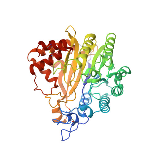
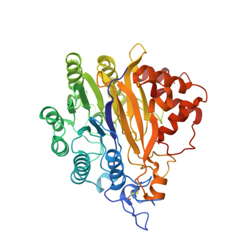
The structure and catalytic mechanism of human sphingomyelin phosphodiesterase like 3a - an acid sphingomyelinase homologue with a novel nucleotide hydrolase activity.

Lim, S.M.Yeung, K.Tresaugues, L.Ling, T.H.Nordlund, P.

(2016) FEBS J 283: 1107-1123

  • DOI: https://doi.org/10.1111/febs.13655
  • Primary Citation Related Structures: 
    2XS6, 5EBB, 5EBE

  • PubMed Abstract: 

    Human sphingomyelinase phosphodiesterase like 3a (SMPDL3a) is a secreted enzyme that shares a conserved catalytic domain with human acid sphingomyelinase (aSMase), the enzyme carrying mutations causative of Niemann-Pick disease. We have solved the structure of SMPDL3a revealing a calcineurin-like fold. A dimetal site, glycosylation pattern and a disulfide bond network are likely to be conserved also in human aSMase. We show that the binuclear site of SMPDL3a is occupied by two Zn(2+) ions and that excess Zn(2+) leads to inhibition of enzyme activity through binding to additional sites. As an extension of recent biochemical work we uncovered that SMPDL3a catalyses the hydrolysis of several modified nucleotides that include cytidine 5'-diphosphocholine, cytidine diphosphate ethanolamine and ADP-ribose, but not the aSMase substrate, sphingomyelin. We subsequently determined the structure of SMPDL3a in complex with the product 5'-cytidine monophosphate (CMP), a structure that is consistent with several distinct coordination modes of the substrate/product in the active site during the reaction cycle. Based on the structure of CMP complexes, we propose a phosphoryl transfer mechanism for SMPDL3a. Finally, a homology model of human aSMase was constructed to allow for the mapping of selected Niemann-Pick disease mutations on a three-dimensional framework to guide further characterization of their effects on aSMase function. Structural data are available in the PDB database under the accession numbers 5EBB and 5EBE.


  • Organizational Affiliation
    • Division of Biomedical Structural Biology, School of Biological Sciences, Nanyang Technological University, Singapore.

Macromolecule Content 

  • Total Structure Weight: 146.51 kDa 
  • Atom Count: 10,472 
  • Modeled Residue Count: 1,248 
  • Deposited Residue Count: 1,248 
  • Unique protein chains: 2

Macromolecules

Find similar proteins by:|  3D Structure
Entity ID: 1
MoleculeChains  Sequence LengthOrganismDetailsImage
Acid sphingomyelinase-like phosphodiesterase 3a418Homo sapiensMutation(s): 0 
Gene Names: SMPDL3AASML3A
EC: 3.1.4 (PDB Primary Data), 3.6.1.15 (UniProt)
Membrane Entity: Yes 
UniProt & NIH Common Fund Data Resources
Find proteins for Q92484 (Homo sapiens)
Explore Q92484 
Go to UniProtKB:  Q92484
PHAROS:  Q92484
GTEx:  ENSG00000172594 
Entity Groups
Sequence Clusters30% Identity50% Identity70% Identity90% Identity95% Identity100% Identity
UniProt GroupQ92484
Glycosylation
Glycosylation Sites: 3Go to GlyGen: Q92484-1
Sequence Annotations
Expand
Reference Sequence
Find similar proteins by:|  3D Structure
Entity ID: 2
MoleculeChains  Sequence LengthOrganismDetailsImage
Acid sphingomyelinase-like phosphodiesterase 3a
B, C
415Homo sapiensMutation(s): 0 
Gene Names: SMPDL3AASML3A
EC: 3.1.4 (PDB Primary Data), 3.6.1.15 (UniProt)
Membrane Entity: Yes 
UniProt & NIH Common Fund Data Resources
Find proteins for Q92484 (Homo sapiens)
Explore Q92484 
Go to UniProtKB:  Q92484
PHAROS:  Q92484
GTEx:  ENSG00000172594 
Entity Groups
Sequence Clusters30% Identity50% Identity70% Identity90% Identity95% Identity100% Identity
UniProt GroupQ92484
Glycosylation
Glycosylation Sites: 4Go to GlyGen: Q92484-1
Sequence Annotations
Expand
Reference Sequence

Oligosaccharides

Help  
Entity ID: 3
MoleculeChains Length2D Diagram GlycosylationD Interactions
2-acetamido-2-deoxy-beta-D-glucopyranose-(1-4)-2-acetamido-2-deoxy-beta-D-glucopyranose
D
2N-Glycosylation
Glycosylation Resources
GlyTouCan: G42666HT
GlyCosmos: G42666HT
GlyGen: G42666HT

Small Molecules

Ligands 7 Unique
IDChains Name / Formula / InChI Key2D Diagram3D Interactions
C5P

Query on C5P


Download:Ideal Coordinates CCD File
AA [auth C],
K [auth A]
CYTIDINE-5'-MONOPHOSPHATE
C9 H14 N3 O8 P
IERHLVCPSMICTF-XVFCMESISA-N
RP5

Query on RP5


Download:Ideal Coordinates CCD File
T [auth B]5-O-phosphono-beta-D-ribofuranose
C5 H11 O8 P
KTVPXOYAKDPRHY-TXICZTDVSA-N
NAG

Query on NAG


Download:Ideal Coordinates CCD File
E [auth A]
F [auth A]
G [auth A]
H [auth A]
L [auth B]
E [auth A],
F [auth A],
G [auth A],
H [auth A],
L [auth B],
M [auth B],
N [auth B],
U [auth C],
V [auth C],
W [auth C],
X [auth C]
2-acetamido-2-deoxy-beta-D-glucopyranose
C8 H15 N O6
OVRNDRQMDRJTHS-FMDGEEDCSA-N
MLI

Query on MLI


Download:Ideal Coordinates CCD File
S [auth B]MALONATE ION
C3 H2 O4
OFOBLEOULBTSOW-UHFFFAOYSA-L
GOL

Query on GOL


Download:Ideal Coordinates CCD File
Q [auth B],
R [auth B]
GLYCEROL
C3 H8 O3
PEDCQBHIVMGVHV-UHFFFAOYSA-N
ZN

Query on ZN


Download:Ideal Coordinates CCD File
I [auth A]
J [auth A]
O [auth B]
P [auth B]
Y [auth C]
I [auth A],
J [auth A],
O [auth B],
P [auth B],
Y [auth C],
Z [auth C]
ZINC ION
Zn
PTFCDOFLOPIGGS-UHFFFAOYSA-N
SCN

Query on SCN


Download:Ideal Coordinates CCD File
BA [auth C],
CA [auth C]
THIOCYANATE ION
C N S
ZMZDMBWJUHKJPS-UHFFFAOYSA-M

Experimental Data & Validation

Experimental Data

  • Method: X-RAY DIFFRACTION
  • Resolution: 3.00 Å
  • R-Value Free:  0.295 (Depositor), 0.294 (DCC) 
  • R-Value Work:  0.251 (Depositor), 0.251 (DCC) 
  • R-Value Observed: 0.254 (Depositor) 
Space Group: P 32 2 1
Unit Cell:
Length ( Å )Angle ( ˚ )
a = 147.654α = 90
b = 147.654β = 90
c = 142.298γ = 120
Software Package:
Software NamePurpose
REFMACrefinement
XDSdata reduction
Aimlessdata scaling
PHASERphasing
Cootmodel building

Structure Validation

View Full Validation Report


Ligand Structure Quality Assessment 


Entry History 

Deposition Data

Revision History  (Full details and data files)

  • Version 1.0: 2016-01-20
    Type: Initial release
  • Version 1.1: 2016-02-03
    Changes: Database references
  • Version 1.2: 2016-03-30
    Changes: Database references
  • Version 2.0: 2020-07-29
    Type: Remediation
    Reason: Carbohydrate remediation
    Changes: Advisory, Atomic model, Data collection, Derived calculations, Structure summary
  • Version 2.1: 2024-01-10
    Changes: Data collection, Database references, Refinement description, Structure summary
  • Version 2.2: 2024-11-20
    Changes: Structure summary