5EAK | pdb_00005eak

Optimization of Microtubule Affinity Regulating Kinase (MARK) Inhibitors with Improved Physical Properties


Experimental Data Snapshot

  • Method: X-RAY DIFFRACTION
  • Resolution: 2.80 Å
  • R-Value Free: 
    0.254 (Depositor), 0.247 (DCC) 
  • R-Value Work: 
    0.195 (Depositor), 0.190 (DCC) 
  • R-Value Observed: 
    0.198 (Depositor) 

wwPDB Validation 3D Report Full Report

Validation slider image for 5EAK

Ligand Structure Quality Assessment 


This is version 1.3 of the entry. See complete history

Literature

Optimization of microtubule affinity regulating kinase (MARK) inhibitors with improved physical properties.

Sloman, D.L.Noucti, N.Altman, M.D.Chen, D.Mislak, A.C.Szewczak, A.Hayashi, M.Warren, L.Dellovade, T.Wu, Z.Marcus, J.Walker, D.Su, H.P.Edavettal, S.C.Munshi, S.Hutton, M.Nuthall, H.Stanton, M.G.

(2016) Bioorg Med Chem Lett 26: 4362-4366

  • DOI: https://doi.org/10.1016/j.bmcl.2016.02.003
  • Primary Citation Related Structures: 
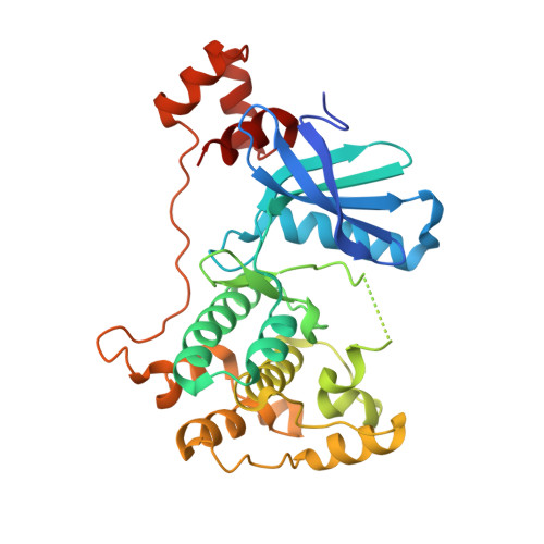
    5EAK

  • PubMed Abstract: 

    Inhibition of microtubule affinity regulating kinase (MARK) represents a potentially attractive means of arresting neurofibrillary tangle pathology in Alzheimer's disease. This manuscript outlines efforts to optimize a pyrazolopyrimidine series of MARK inhibitors by focusing on improvements in potency, physical properties and attributes amenable to CNS penetration. A unique cylcyclohexyldiamine scaffold was identified that led to remarkable improvements in potency, opening up opportunities to reduce MW, Pgp efflux and improve pharmacokinetic properties while also conferring improved solubility.


  • Organizational Affiliation
    • Discovery Chemistry, Merck and Co., Inc., 33 Avenue Louis Pasteur, Boston, MA 02215, United States.

Macromolecule Content 

  • Total Structure Weight: 76.51 kDa 
  • Atom Count: 4,981 
  • Modeled Residue Count: 605 
  • Deposited Residue Count: 656 
  • Unique protein chains: 1

Macromolecules

Find similar proteins by:|  3D Structure
Entity ID: 1
MoleculeChains  Sequence LengthOrganismDetailsImage
Serine/threonine-protein kinase MARK2
A, B
328Homo sapiensMutation(s): 0 
Gene Names: MARK2EMK1
EC: 2.7.11.1 (PDB Primary Data), 2.7.11.26 (PDB Primary Data)
UniProt & NIH Common Fund Data Resources
Find proteins for Q7KZI7 (Homo sapiens)
Explore Q7KZI7 
Go to UniProtKB:  Q7KZI7
PHAROS:  Q7KZI7
GTEx:  ENSG00000072518 
Entity Groups
Sequence Clusters30% Identity50% Identity70% Identity90% Identity95% Identity100% Identity
UniProt GroupQ7KZI7
Sequence Annotations
Expand
Reference Sequence

Small Molecules

Ligands 1 Unique
IDChains Name / Formula / InChI Key2D Diagram3D Interactions
24R

Query on 24R


Download:Ideal Coordinates CCD File
C [auth A],
D [auth B]
N-[(1S,2R)-2-aminocyclohexyl]-4-[6-(1-methyl-1H-pyrazol-4-yl)pyrazolo[1,5-a]pyrimidin-3-yl]thiophene-2-carboxamide
C21 H23 N7 O S
LYADGAGFYYXXIO-MSOLQXFVSA-N

Experimental Data & Validation

Experimental Data

  • Method: X-RAY DIFFRACTION
  • Resolution: 2.80 Å
  • R-Value Free:  0.254 (Depositor), 0.247 (DCC) 
  • R-Value Work:  0.195 (Depositor), 0.190 (DCC) 
  • R-Value Observed: 0.198 (Depositor) 
Space Group: P 61
Unit Cell:
Length ( Å )Angle ( ˚ )
a = 118.739α = 90
b = 118.739β = 90
c = 106.592γ = 120
Software Package:
Software NamePurpose
HKL-2000data reduction
HKL-2000data scaling
REFMACphasing
PHENIXrefinement
DENZOdata reduction
SCALEPACKdata scaling

Structure Validation

View Full Validation Report


Ligand Structure Quality Assessment 


Entry History 

Deposition Data

  • Released Date: 2016-02-17 
  • Deposition Author(s): Su, H.P.

Revision History  (Full details and data files)

  • Version 1.0: 2016-02-17
    Type: Initial release
  • Version 1.1: 2016-08-24
    Changes: Database references
  • Version 1.2: 2017-11-22
    Changes: Database references, Derived calculations, Refinement description
  • Version 1.3: 2024-03-06
    Changes: Data collection, Database references