5E8J | pdb_00005e8j

Crystal structure of mRNA cap guanine-N7 methyltransferase in complex with RAM


Experimental Data Snapshot

  • Method: X-RAY DIFFRACTION
  • Resolution: 2.35 Å
  • R-Value Free: 
    0.269 (Depositor), 0.270 (DCC) 
  • R-Value Work: 
    0.210 (Depositor), 0.210 (DCC) 
  • R-Value Observed: 
    0.213 (Depositor) 

Starting Model: experimental
View more details

wwPDB Validation   3D Report Full Report


Ligand Structure Quality Assessment 

Created with Raphaël 2.3.0Worse 01 BetterLigand structure goodness of fit to experimental dataBest fitted SAHClick on this verticalbar to view details

This is version 1.5 of the entry. See complete history


Literature

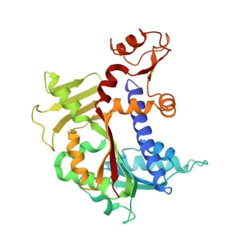
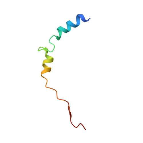
Molecular basis of RNA guanine-7 methyltransferase (RNMT) activation by RAM.

Varshney, D.Petit, A.P.Bueren-Calabuig, J.A.Jansen, C.Fletcher, D.A.Peggie, M.Weidlich, S.Scullion, P.Pisliakov, A.V.Cowling, V.H.

(2016) Nucleic Acids Res 44: 10423-10436

  • DOI: https://doi.org/10.1093/nar/gkw637
  • Primary Citation of Related Structures:  
    5E8J, 5E9J, 5E9W

  • PubMed Abstract: 

    Maturation and translation of mRNA in eukaryotes requires the addition of the 7-methylguanosine cap. In vertebrates, the cap methyltransferase, RNA guanine-7 methyltransferase (RNMT), has an activating subunit, RNMT-Activating Miniprotein (RAM). Here we report the first crystal structure of the human RNMT in complex with the activation domain of RAM. A relatively unstructured and negatively charged RAM binds to a positively charged surface groove on RNMT, distal to the active site. This results in stabilisation of a RNMT lobe structure which co-evolved with RAM and is required for RAM binding. Structure-guided mutagenesis and molecular dynamics simulations reveal that RAM stabilises the structure and positioning of the RNMT lobe and the adjacent α-helix hinge, resulting in optimal positioning of helix A which contacts substrates in the active site. Using biophysical and biochemical approaches, we observe that RAM increases the recruitment of the methyl donor, AdoMet (S-adenosyl methionine), to RNMT. Thus we report the mechanism by which RAM allosterically activates RNMT, allowing it to function as a molecular rheostat for mRNA cap methylation.


  • Organizational Affiliation

    Centre for Gene Regulation and Expression, School of Life Sciences, University of Dundee, Dow Street, Dundee DD1 5EH, UK.


Macromolecules
Find similar proteins by:  (by identity cutoff)  |  3D Structure
Entity ID: 1
MoleculeChains Sequence LengthOrganismDetailsImage
mRNA cap guanine-N7 methyltransferase
A, B
312Homo sapiensMutation(s): 0 
Gene Names: RNMTKIAA0398
EC: 2.1.1.56
UniProt & NIH Common Fund Data Resources
Find proteins for O43148 (Homo sapiens)
Explore O43148 
Go to UniProtKB:  O43148
PHAROS:  O43148
GTEx:  ENSG00000101654 
Entity Groups  
Sequence Clusters30% Identity50% Identity70% Identity90% Identity95% Identity100% Identity
UniProt GroupO43148
Sequence Annotations
Expand
  • Reference Sequence
Find similar proteins by:  (by identity cutoff)  |  3D Structure
Entity ID: 2
MoleculeChains Sequence LengthOrganismDetailsImage
RNMT-activating mini protein
C, D
44Homo sapiensMutation(s): 0 
Gene Names: FAM103A1C15orf18
UniProt & NIH Common Fund Data Resources
Find proteins for Q9BTL3 (Homo sapiens)
Explore Q9BTL3 
Go to UniProtKB:  Q9BTL3
PHAROS:  Q9BTL3
GTEx:  ENSG00000169612 
Entity Groups  
Sequence Clusters30% Identity50% Identity70% Identity90% Identity95% Identity100% Identity
UniProt GroupQ9BTL3
Sequence Annotations
Expand
  • Reference Sequence
Experimental Data & Validation

Experimental Data

  • Method: X-RAY DIFFRACTION
  • Resolution: 2.35 Å
  • R-Value Free:  0.269 (Depositor), 0.270 (DCC) 
  • R-Value Work:  0.210 (Depositor), 0.210 (DCC) 
  • R-Value Observed: 0.213 (Depositor) 
Space Group: P 1
Unit Cell:
Length ( Å )Angle ( ˚ )
a = 49.147α = 90.06
b = 50.487β = 92.41
c = 84.579γ = 115.41
Software Package:
Software NamePurpose
SCALAdata scaling
MOLREPphasing
PHENIXrefinement
PDB_EXTRACTdata extraction
XDSdata reduction

Structure Validation

View Full Validation Report


Ligand Structure Quality Assessment 

Created with Raphaël 2.3.0Worse 01 BetterLigand structure goodness of fit to experimental dataBest fitted SAHClick on this verticalbar to view details

Entry History 

Deposition Data

Revision History  (Full details and data files)

  • Version 1.0: 2016-07-13
    Type: Initial release
  • Version 1.1: 2016-08-03
    Changes: Database references
  • Version 1.2: 2016-12-14
    Changes: Database references
  • Version 1.3: 2019-02-20
    Changes: Advisory, Data collection, Derived calculations
  • Version 1.4: 2019-07-10
    Changes: Data collection
  • Version 1.5: 2024-01-10
    Changes: Data collection, Database references, Refinement description