5E80 | pdb_00005e80

The crystal structure of PDEd in complex with inhibitor-2a


Experimental Data Snapshot

  • Method: X-RAY DIFFRACTION
  • Resolution: 2.60 Å
  • R-Value Free: 
    0.247 (Depositor), 0.245 (DCC) 
  • R-Value Work: 
    0.185 (Depositor), 0.189 (DCC) 
  • R-Value Observed: 
    0.188 (Depositor) 

Starting Model: experimental
View more details

wwPDB Validation 3D Report Full Report

Validation slider image for 5E80

Ligand Structure Quality Assessment 


This is version 1.1 of the entry. See complete history

Literature

Identification of pyrazolopyridazinones as PDE delta inhibitors.

Papke, B.Murarka, S.Vogel, H.A.Martin-Gago, P.Kovacevic, M.Truxius, D.C.Fansa, E.K.Ismail, S.Zimmermann, G.Heinelt, K.Schultz-Fademrecht, C.Al Saabi, A.Baumann, M.Nussbaumer, P.Wittinghofer, A.Waldmann, H.Bastiaens, P.I.

(2016) Nat Commun 7: 11360-11360

  • DOI: https://doi.org/10.1038/ncomms11360
  • Primary Citation Related Structures: 
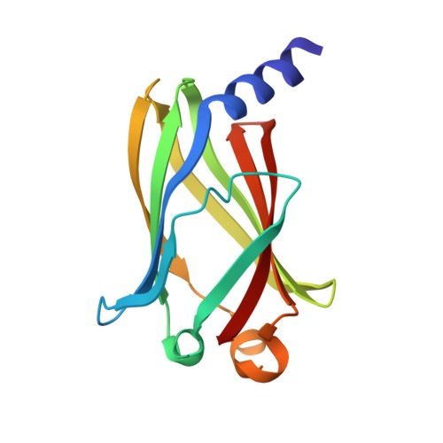
    5E80

  • PubMed Abstract: 

    The prenyl-binding protein PDEδ is crucial for the plasma membrane localization of prenylated Ras. Recently, we have reported that the small-molecule Deltarasin binds to the prenyl-binding pocket of PDEδ, and impairs Ras enrichment at the plasma membrane, thereby affecting the proliferation of KRas-dependent human pancreatic ductal adenocarcinoma cell lines. Here, using structure-based compound design, we have now identified pyrazolopyridazinones as a novel, unrelated chemotype that binds to the prenyl-binding pocket of PDEδ with high affinity, thereby displacing prenylated Ras proteins in cells. Our results show that the new PDEδ inhibitor, named Deltazinone 1, is highly selective, exhibits less unspecific cytotoxicity than the previously reported Deltarasin and demonstrates a high correlation with the phenotypic effect of PDEδ knockdown in a set of human pancreatic cancer cell lines.


  • Organizational Affiliation
    • Department of Systemic Cell Biology, Max Planck Institute of Molecular Physiology, D-44227 Dortmund, Germany.

Macromolecule Content 

  • Total Structure Weight: 35.4 kDa 
  • Atom Count: 2,503 
  • Modeled Residue Count: 298 
  • Deposited Residue Count: 298 
  • Unique protein chains: 1

Macromolecules

Find similar proteins by:|  3D Structure
Entity ID: 1
MoleculeChains  Sequence LengthOrganismDetailsImage
Retinal rod rhodopsin-sensitive cGMP 3',5'-cyclic phosphodiesterase subunit deltaA [auth B],
B [auth A]
149Homo sapiensMutation(s): 0 
Gene Names: PDE6DPDED
UniProt & NIH Common Fund Data Resources
Find proteins for O43924 (Homo sapiens)
Explore O43924 
Go to UniProtKB:  O43924
PHAROS:  O43924
GTEx:  ENSG00000156973 
Entity Groups
Sequence Clusters30% Identity50% Identity70% Identity90% Identity95% Identity100% Identity
UniProt GroupO43924
Sequence Annotations
Expand
Reference Sequence

Small Molecules

Ligands 1 Unique
IDChains Name / Formula / InChI Key2D Diagram3D Interactions
5KP

Query on 5KP


Download:Ideal Coordinates CCD File
C [auth B],
D [auth A]
N-(3-chloro-2-methylphenyl)-4-(3,4-dimethyl-7-oxo-2-phenyl-2,7-dihydro-6H-pyrazolo[3,4-d]pyridazin-6-yl)butanamide
C24 H24 Cl N5 O2
HPMUIOXFVVIJSJ-UHFFFAOYSA-N

Experimental Data & Validation

Experimental Data

  • Method: X-RAY DIFFRACTION
  • Resolution: 2.60 Å
  • R-Value Free:  0.247 (Depositor), 0.245 (DCC) 
  • R-Value Work:  0.185 (Depositor), 0.189 (DCC) 
  • R-Value Observed: 0.188 (Depositor) 
Space Group: P 1
Unit Cell:
Length ( Å )Angle ( ˚ )
a = 31.76α = 97.7
b = 40.9β = 102.38
c = 68.8γ = 89.31
Software Package:
Software NamePurpose
XDSdata reduction
XSCALEdata scaling
MOLREPphasing
REFMACrefinement
Cootmodel building

Structure Validation

View Full Validation Report


Ligand Structure Quality Assessment 


Entry History 

Deposition Data

Revision History  (Full details and data files)

  • Version 1.0: 2016-05-04
    Type: Initial release
  • Version 1.1: 2024-01-10
    Changes: Data collection, Database references, Refinement description