5E7S | pdb_00005e7s

Hexameric structure of a LonA protease domain in active state


Experimental Data Snapshot

  • Method: X-RAY DIFFRACTION
  • Resolution: 3.03 Å
  • R-Value Free: 
    0.279 (Depositor), 0.257 (DCC) 
  • R-Value Work: 
    0.227 (Depositor) 
  • R-Value Observed: 
    0.230 (Depositor) 

Starting Model: experimental
View more details

wwPDB Validation 3D Report Full Report

Validation slider image for 5E7S

This is version 1.3 of the entry. See complete history

Literature

Structural Basis for the Magnesium-Dependent Activation and Hexamerization of the Lon AAA+ Protease

Su, S.-C.Lin, C.-C.Tai, H.-C.Chang, M.-Y.Ho, M.-R.Babu, C.S.Liao, J.-H.Wu, S.-H.Chang, Y.-C.Lim, C.Chang, C.-I.

(2016) Structure 24: 676-686

  • DOI: https://doi.org/10.1016/j.str.2016.03.003
  • Primary Citation Related Structures: 
    4YPM, 5E7S

  • PubMed Abstract: 

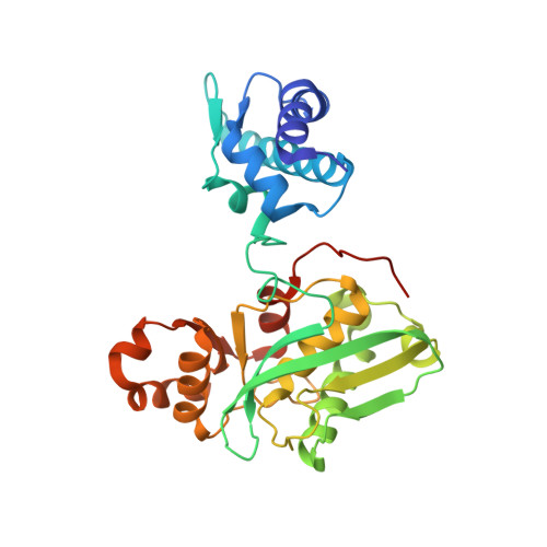
    The Lon AAA+ protease (LonA) plays important roles in protein homeostasis and regulation of diverse biological processes. LonA behaves as a homomeric hexamer in the presence of magnesium (Mg(2+)) and performs ATP-dependent proteolysis. However, it is also found that LonA can carry out Mg(2+)-dependent degradation of unfolded protein substrate in an ATP-independent manner. Here we show that in the presence of Mg(2+) LonA forms a non-secluded hexameric barrel with prominent openings, which explains why Mg(2+)-activated LonA can operate as a diffusion-based chambered protease to degrade unstructured protein and peptide substrates efficiently in the absence of ATP. A 1.85 Å crystal structure of Mg(2+)-activated protease domain reveals Mg(2+)-dependent remodeling of a substrate-binding loop and a potential metal-binding site near the Ser-Lys catalytic dyad, supported by biophysical binding assays and molecular dynamics simulations. Together, these findings reveal the specific roles of Mg(2+) in the molecular assembly and activation of LonA.


  • Organizational Affiliation
    • Institute of Biological Chemistry, Academia Sinica, Taipei, Taiwan 11529, ROC; Institute of Biochemical Sciences, National Taiwan University, Taipei, Taiwan 10617, ROC.

Macromolecule Content 

  • Total Structure Weight: 391.25 kDa 
  • Atom Count: 26,298 
  • Modeled Residue Count: 3,443 
  • Deposited Residue Count: 3,576 
  • Unique protein chains: 1

Macromolecules

Find similar proteins by:|  3D Structure
Entity ID: 1
MoleculeChains  Sequence LengthOrganismDetailsImage
Lon protease
A, B, C, D, E
A, B, C, D, E, F, G, H, I, J, K, L
298Meiothermus taiwanensisMutation(s): 0 
Gene Names: lonA1lon
EC: 3.4.21.53
UniProt
Find proteins for A0A059VAZ3 (Meiothermus taiwanensis)
Explore A0A059VAZ3 
Go to UniProtKB:  A0A059VAZ3
Entity Groups
Sequence Clusters30% Identity50% Identity70% Identity90% Identity95% Identity100% Identity
UniProt GroupA0A059VAZ3
Sequence Annotations
Expand
Reference Sequence

Experimental Data & Validation

Experimental Data

  • Method: X-RAY DIFFRACTION
  • Resolution: 3.03 Å
  • R-Value Free:  0.279 (Depositor), 0.257 (DCC) 
  • R-Value Work:  0.227 (Depositor) 
  • R-Value Observed: 0.230 (Depositor) 
Space Group: C 1 2 1
Unit Cell:
Length ( Å )Angle ( ˚ )
a = 116.309α = 90
b = 212.787β = 100.8
c = 178.921γ = 90
Software Package:
Software NamePurpose
REFMACrefinement
HKL-2000data reduction
HKL-2000data scaling
Cootmodel building

Structure Validation

View Full Validation Report


Entry History 

Deposition Data

Revision History  (Full details and data files)

  • Version 1.0: 2016-03-16
    Type: Initial release
  • Version 1.1: 2016-04-20
    Changes: Database references
  • Version 1.2: 2016-05-18
    Changes: Database references, Experimental preparation
  • Version 1.3: 2023-11-08
    Changes: Data collection, Database references, Derived calculations, Refinement description