5E6T | pdb_00005e6t

Crystal structure of bovine norovirus P domain


Experimental Data Snapshot

  • Method: X-RAY DIFFRACTION
  • Resolution: 2.27 Å
  • R-Value Free: 
    0.241 (Depositor), 0.242 (DCC) 
  • R-Value Work: 
    0.195 (Depositor), 0.197 (DCC) 
  • R-Value Observed: 
    0.197 (Depositor) 

Starting Model: experimental
View more details

wwPDB Validation 3D Report Full Report

Validation slider image for 5E6T

This is version 1.1 of the entry. See complete history

Literature

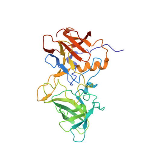
Structural analysis of bovine norovirus protruding domain.

Singh, B.K.Koromyslova, A.Hansman, G.S.

(2015) Virology 487: 296-301

  • DOI: https://doi.org/10.1016/j.virol.2015.10.022
  • Primary Citation Related Structures: 
    5E6T

  • PubMed Abstract: 

    We determined a structure of a bovine (genogroup III, GIII) norovirus capsid protruding (P) domain using X-ray crystallography. The bovine P domain was reminiscent of other norovirus genogroups (GI, GII, GIV, and GV), but closely matched the human GI P domain. We also identified a monoclonal antibody that was capable of binding the five different (GI-GV) P domains. Our data suggests that genetically diverse noroviruses still contain common epitopes.


  • Organizational Affiliation
    • Schaller Research Group at the University of Heidelberg and the DKFZ, Heidelberg 69120, Germany; Department of Infectious Diseases, Virology, University of Heidelberg, Heidelberg 69120, Germany.

Macromolecule Content 

  • Total Structure Weight: 63.24 kDa 
  • Atom Count: 4,614 
  • Modeled Residue Count: 572 
  • Deposited Residue Count: 576 
  • Unique protein chains: 1

Macromolecules

Find similar proteins by:|  3D Structure
Entity ID: 1
MoleculeChains  Sequence LengthOrganismDetailsImage
Capsid
A, B
288Norovirus Bo/GIII/B309/2003/BELMutation(s): 0 
UniProt
Find proteins for E6Y5B1 (Norovirus Bo/GIII/B309/2003/BEL)
Explore E6Y5B1 
Go to UniProtKB:  E6Y5B1
Entity Groups
Sequence Clusters30% Identity50% Identity70% Identity90% Identity95% Identity100% Identity
UniProt GroupE6Y5B1
Sequence Annotations
Expand
Reference Sequence

Experimental Data & Validation

Experimental Data

  • Method: X-RAY DIFFRACTION
  • Resolution: 2.27 Å
  • R-Value Free:  0.241 (Depositor), 0.242 (DCC) 
  • R-Value Work:  0.195 (Depositor), 0.197 (DCC) 
  • R-Value Observed: 0.197 (Depositor) 
Space Group: C 1 2 1
Unit Cell:
Length ( Å )Angle ( ˚ )
a = 112.92α = 90
b = 63.86β = 115.06
c = 87.56γ = 90
Software Package:
Software NamePurpose
XSCALEdata scaling
PHASERphasing
PHENIXrefinement
PDB_EXTRACTdata extraction
XDSdata reduction

Structure Validation

View Full Validation Report


Entry History 

& Funding Information

Deposition Data


Funding OrganizationLocationGrant Number
CHS foundationGermany--
Chinese Academy of SciencesChina--

Revision History  (Full details and data files)

  • Version 1.0: 2015-12-09
    Type: Initial release
  • Version 1.1: 2024-01-10
    Changes: Data collection, Database references, Derived calculations, Refinement description