5E4R | pdb_00005e4r

Crystal structure of domain-duplicated synthetic class II ketol-acid reductoisomerase 2Ia_KARI-DD


Experimental Data Snapshot

  • Method: X-RAY DIFFRACTION
  • Resolution: 1.94 Å
  • R-Value Free: 
    0.179 (Depositor), 0.191 (DCC) 
  • R-Value Work: 
    0.158 (Depositor), 0.168 (DCC) 
  • R-Value Observed: 
    0.159 (Depositor) 

Starting Model: experimental
View more details

wwPDB Validation 3D Report Full Report

Validation slider image for 5E4R

Ligand Structure Quality Assessment 


This is version 1.3 of the entry. See complete history

Literature

Artificial domain duplication replicates evolutionary history of ketol-acid reductoisomerases.

Cahn, J.K.Brinkmann-Chen, S.Buller, A.R.Arnold, F.H.

(2016) Protein Sci 25: 1241-1248

  • DOI: https://doi.org/10.1002/pro.2852
  • Primary Citation Related Structures: 
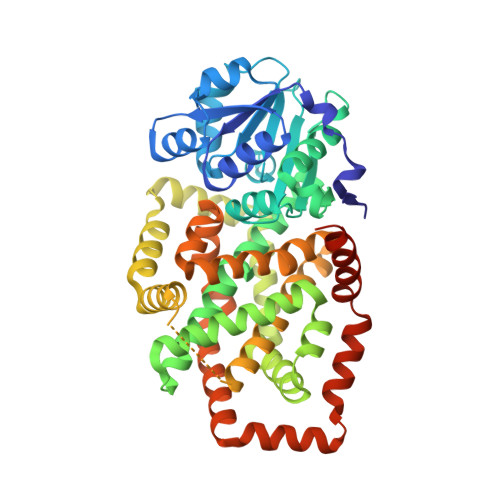
    5E4R

  • PubMed Abstract: 

    The duplication of protein structural domains has been proposed as a common mechanism for the generation of new protein folds. A particularly interesting case is the class II ketol-acid reductoisomerase (KARI), which putatively arose from an ancestral class I KARI by duplication of the C-terminal domain and corresponding loss of obligate dimerization. As a result, the class II enzymes acquired a deeply embedded figure-of-eight knot. To test this evolutionary hypothesis we constructed a novel class II KARI by duplicating the C-terminal domain of a hyperthermostable class I KARI. The new protein is monomeric, as confirmed by gel filtration and X-ray crystallography, and has the deeply knotted class II KARI fold. Surprisingly, its catalytic activity is nearly unchanged from the parent KARI. This provides strong evidence in support of domain duplication as the mechanism for the evolution of the class II KARI fold and demonstrates the ability of domain duplication to generate topological novelty in a function-neutral manner.


  • Organizational Affiliation
    • Division of Chemistry and Chemical Engineering, California Institute of Technology, Pasadena, California, 91125.

Macromolecule Content 

  • Total Structure Weight: 56.34 kDa 
  • Atom Count: 4,060 
  • Modeled Residue Count: 466 
  • Deposited Residue Count: 490 
  • Unique protein chains: 1

Macromolecules

Find similar proteins by:|  3D Structure
Entity ID: 1
MoleculeChains  Sequence LengthOrganismDetailsImage
Ketol-acid reductoisomerase490Ignisphaera aggregans DSM 17230Mutation(s): 0 
Gene Names: ilvCIgag_1561
EC: 1.1.1.86 (PDB Primary Data), 1.1.1.383 (UniProt)
UniProt
Find proteins for E0SRA9 (Ignisphaera aggregans (strain DSM 17230 / JCM 13409 / AQ1.S1))
Explore E0SRA9 
Go to UniProtKB:  E0SRA9
Entity Groups
Sequence Clusters30% Identity50% Identity70% Identity90% Identity95% Identity100% Identity
UniProt GroupE0SRA9
Sequence Annotations
Expand
Reference Sequence

Small Molecules

Ligands 5 Unique
IDChains Name / Formula / InChI Key2D Diagram3D Interactions
NAP

Query on NAP


Download:Ideal Coordinates CCD File
B [auth A]NADP NICOTINAMIDE-ADENINE-DINUCLEOTIDE PHOSPHATE
C21 H28 N7 O17 P3
XJLXINKUBYWONI-NNYOXOHSSA-N
40E

Query on 40E


Download:Ideal Coordinates CCD File
E [auth A]oxo(propan-2-ylamino)acetic acid
C5 H9 N O3
KBMFHCMLHQGQEB-UHFFFAOYSA-N
GOL

Query on GOL


Download:Ideal Coordinates CCD File
G [auth A]GLYCEROL
C3 H8 O3
PEDCQBHIVMGVHV-UHFFFAOYSA-N
EDO

Query on EDO


Download:Ideal Coordinates CCD File
F [auth A]1,2-ETHANEDIOL
C2 H6 O2
LYCAIKOWRPUZTN-UHFFFAOYSA-N
MG

Query on MG


Download:Ideal Coordinates CCD File
C [auth A],
D [auth A]
MAGNESIUM ION
Mg
JLVVSXFLKOJNIY-UHFFFAOYSA-N

Experimental Data & Validation

Experimental Data

  • Method: X-RAY DIFFRACTION
  • Resolution: 1.94 Å
  • R-Value Free:  0.179 (Depositor), 0.191 (DCC) 
  • R-Value Work:  0.158 (Depositor), 0.168 (DCC) 
  • R-Value Observed: 0.159 (Depositor) 
Space Group: P 43 2 2
Unit Cell:
Length ( Å )Angle ( ˚ )
a = 103.882α = 90
b = 103.882β = 90
c = 142.074γ = 90
Software Package:
Software NamePurpose
REFMACrefinement
SCALAdata scaling
PHASERphasing
PDB_EXTRACTdata extraction
XDSdata reduction

Structure Validation

View Full Validation Report


Ligand Structure Quality Assessment 


Entry History 

& Funding Information

Deposition Data


Funding OrganizationLocationGrant Number
Gordon and Betty Moore FoundationUnited StatesGBMF2809

Revision History  (Full details and data files)

  • Version 1.0: 2015-12-23
    Type: Initial release
  • Version 1.1: 2015-12-30
    Changes: Database references
  • Version 1.2: 2016-07-06
    Changes: Database references
  • Version 1.3: 2023-09-27
    Changes: Data collection, Database references, Derived calculations, Refinement description