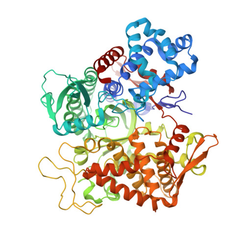
5E2Q

Structure of human DPP3 in complex with angiotensin-II


Experimental Data Snapshot

  • Method: X-RAY DIFFRACTION
  • Resolution: 2.40 Å
  • R-Value Free: 0.247 
  • R-Value Work: 0.206 
  • R-Value Observed: 0.208 

Starting Model: experimental
View more details

wwPDB Validation   3D Report Full Report


This is version 2.0 of the entry. See complete history


Literature

Substrate complexes of human dipeptidyl peptidase III reveal the mechanism of enzyme inhibition.

Kumar, P.Reithofer, V.Reisinger, M.Wallner, S.Pavkov-Keller, T.Macheroux, P.Gruber, K.

(2016) Sci Rep 6: 23787-23787

  • DOI: https://doi.org/10.1038/srep23787
  • Primary Citation of Related Structures:  
    5E2Q, 5E33, 5E3A, 5E3C, 5EGY, 5EHH

  • PubMed Abstract: 

    Human dipeptidyl-peptidase III (hDPP III) is a zinc-dependent hydrolase cleaving dipeptides off the N-termini of various bioactive peptides. Thus, the enzyme is likely involved in a number of physiological processes such as nociception and is also implicated in several forms of cancer. We present high-resolution crystal structures of hDPP III in complex with opioid peptides (Met-and Leu-enkephalin, endomorphin-2) as well as with angiotensin-II and the peptide inhibitor IVYPW. These structures confirm the previously reported large conformational change of the enzyme upon ligand binding and show that the structure of the closed conformation is independent of the nature of the bound peptide. The overall peptide-binding mode is also conserved ensuring the correct positioning of the scissile peptide bond with respect to the catalytic zinc ion. The structure of the angiotensin-II complex shows, how longer peptides are accommodated in the binding cleft of hDPP III. Differences in the binding modes allow a distinction between real substrates and inhibitory peptides or "slow" substrates. The latter displace a zinc bound water molecule necessitating the energetically much less favoured anhydride mechanism as opposed to the favoured promoted-water mechanism. The structural data also form the necessary framework for the design of specific hDPP III inhibitors.


  • Organizational Affiliation

    Institute of Molecular Biosciences, University of Graz, Humboldtstraße 50/3, 8010 Graz, Austria.


Macromolecules
Find similar proteins by:  (by identity cutoff)  |  3D Structure
Entity ID: 1
MoleculeChains Sequence LengthOrganismDetailsImage
Dipeptidyl peptidase 3726Homo sapiensMutation(s): 6 
Gene Names: DPP3
EC: 3.4.14.4
UniProt & NIH Common Fund Data Resources
Find proteins for Q9NY33 (Homo sapiens)
Explore Q9NY33 
Go to UniProtKB:  Q9NY33
PHAROS:  Q9NY33
GTEx:  ENSG00000254986 
Entity Groups  
Sequence Clusters30% Identity50% Identity70% Identity90% Identity95% Identity100% Identity
UniProt GroupQ9NY33
Sequence Annotations
Expand
  • Reference Sequence

Find similar proteins by:  Sequence   |   3D Structure  

Entity ID: 2
MoleculeChains Sequence LengthOrganismDetailsImage
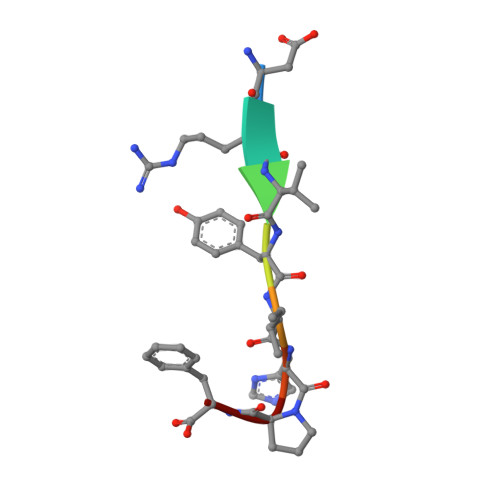
angiotensin-II8Homo sapiensMutation(s): 0 
UniProt & NIH Common Fund Data Resources
Find proteins for P01019 (Homo sapiens)
Explore P01019 
Go to UniProtKB:  P01019
PHAROS:  P01019
GTEx:  ENSG00000135744 
Entity Groups  
UniProt GroupP01019
Sequence Annotations
Expand
  • Reference Sequence
Small Molecules
Ligands 2 Unique
IDChains Name / Formula / InChI Key2D Diagram3D Interactions
K
Query on K

Download Ideal Coordinates CCD File 
D [auth A]POTASSIUM ION
K
NPYPAHLBTDXSSS-UHFFFAOYSA-N
MG
Query on MG

Download Ideal Coordinates CCD File 
C [auth A]MAGNESIUM ION
Mg
JLVVSXFLKOJNIY-UHFFFAOYSA-N
Experimental Data & Validation

Experimental Data

  • Method: X-RAY DIFFRACTION
  • Resolution: 2.40 Å
  • R-Value Free: 0.247 
  • R-Value Work: 0.206 
  • R-Value Observed: 0.208 
  • Space Group: C 1 2 1
Unit Cell:
Length ( Å )Angle ( ˚ )
a = 119.131α = 90
b = 105.922β = 93.91
c = 64.846γ = 90
Software Package:
Software NamePurpose
PHENIXrefinement
XDSdata reduction
SCALAdata scaling
PHENIXphasing

Structure Validation

View Full Validation Report


Entry History & Funding Information

Deposition Data


Funding OrganizationLocationGrant Number
Austrian Science FundAustriaW901

Revision History  (Full details and data files)

  • Version 1.0: 2016-04-13
    Type: Initial release
  • Version 1.1: 2017-09-06
    Changes: Author supporting evidence
  • Version 2.0: 2024-01-10
    Changes: Atomic model, Data collection, Database references, Refinement description