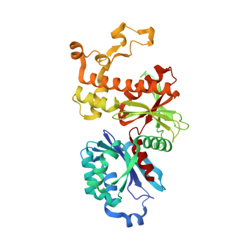
Crystal structure of human PANK2: the catalytic core domain in complex with pantothenate and adenosine diphosphate
LOPPNAU, P., DONG, A., RAVICHANDRAN, M., CHENG, C., TEMPEL, W., SEITOVA, A., HUTCHINSON, A., HONG, B.S., Bountra, C., Arrowsmith, C.H., Edwards, A.M., BROWN, P.J., Structural Genomics Consortium (SGC)To be published.