5E1E | pdb_00005e1e

Human JAK1 kinase in complex with compound 30 at 2.30 Angstroms resolution


Experimental Data Snapshot

  • Method: X-RAY DIFFRACTION
  • Resolution: 2.30 Å
  • R-Value Free: 
    0.268 (Depositor), 0.267 (DCC) 
  • R-Value Work: 
    0.225 (Depositor), 0.219 (DCC) 
  • R-Value Observed: 
    0.227 (Depositor) 

wwPDB Validation 3D Report Full Report

Validation slider image for 5E1E

Ligand Structure Quality Assessment 


This is version 1.3 of the entry. See complete history

Literature

Identification of azabenzimidazoles as potent JAK1 selective inhibitors.

Vasbinder, M.M.Alimzhanov, M.Augustin, M.Bebernitz, G.Bell, K.Chuaqui, C.Deegan, T.Ferguson, A.D.Goodwin, K.Huszar, D.Kawatkar, A.Kawatkar, S.Read, J.Shi, J.Steinbacher, S.Steuber, H.Su, Q.Toader, D.Wang, H.Woessner, R.Wu, A.Ye, M.Zinda, M.

(2016) Bioorg Med Chem Lett 26: 60-67

  • DOI: https://doi.org/10.1016/j.bmcl.2015.11.031
  • Primary Citation Related Structures: 
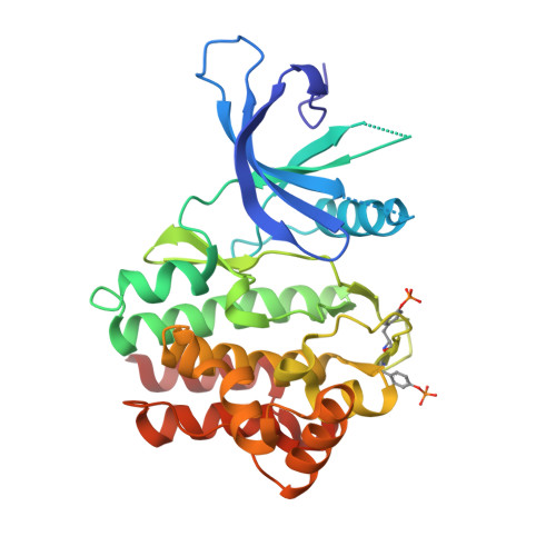
    5E1E

  • PubMed Abstract: 

    We have identified a class of azabenzimidazoles as potent and selective JAK1 inhibitors. Investigations into the SAR are presented along with the structural features required to achieve selectivity for JAK1 versus other JAK family members. An example from the series demonstrated highly selective inhibition of JAK1 versus JAK2 and JAK3, along with inhibition of pSTAT3 in vivo, enabling it to serve as a JAK1 selective tool compound to further probe the biology of JAK1 selective inhibitors.


  • Organizational Affiliation
    • AstraZeneca R&D Boston, Oncology IMED, 35 Gatehouse Drive, Waltham, MA 02451, United States. Electronic address: melissa.vasbinder@astrazeneca.com.

Macromolecule Content 

  • Total Structure Weight: 67.92 kDa 
  • Atom Count: 4,944 
  • Modeled Residue Count: 563 
  • Deposited Residue Count: 580 
  • Unique protein chains: 1

Macromolecules

Find similar proteins by:|  3D Structure
Entity ID: 1
MoleculeChains  Sequence LengthOrganismDetailsImage
Tyrosine-protein kinase JAK1
A, B
290Homo sapiensMutation(s): 0 
Gene Names: JAK1JAK1AJAK1B
EC: 2.7.10.2
UniProt & NIH Common Fund Data Resources
Find proteins for P23458 (Homo sapiens)
Explore P23458 
Go to UniProtKB:  P23458
PHAROS:  P23458
GTEx:  ENSG00000162434 
Entity Groups
Sequence Clusters30% Identity50% Identity70% Identity90% Identity95% Identity100% Identity
UniProt GroupP23458
Sequence Annotations
Expand
Reference Sequence

Small Molecules

Ligands 2 Unique
IDChains Name / Formula / InChI Key2D Diagram3D Interactions
5JG

Query on 5JG


Download:Ideal Coordinates CCD File
D [auth A],
E [auth B]
6-chloro-2-(2-fluoro-4,5-dimethoxyphenyl)-N-(piperidin-4-ylmethyl)-3H-imidazo[4,5-b]pyridin-7-amine
C20 H23 Cl F N5 O2
IRGAIDAWHGYOKD-UHFFFAOYSA-N
PEG

Query on PEG


Download:Ideal Coordinates CCD File
C [auth A]DI(HYDROXYETHYL)ETHER
C4 H10 O3
MTHSVFCYNBDYFN-UHFFFAOYSA-N
Modified Residues  1 Unique
IDChains TypeFormula2D DiagramParent
PTR
Query on PTR
A, B
L-PEPTIDE LINKINGC9 H12 N O6 PTYR

Experimental Data & Validation

Experimental Data

  • Method: X-RAY DIFFRACTION
  • Resolution: 2.30 Å
  • R-Value Free:  0.268 (Depositor), 0.267 (DCC) 
  • R-Value Work:  0.225 (Depositor), 0.219 (DCC) 
  • R-Value Observed: 0.227 (Depositor) 
Space Group: P 21 21 21
Unit Cell:
Length ( Å )Angle ( ˚ )
a = 43.095α = 90
b = 88.708β = 90
c = 174.928γ = 90
Software Package:
Software NamePurpose
BUSTERrefinement
XSCALEdata scaling
XDSdata reduction
MOLREPphasing
Cootmodel building

Structure Validation

View Full Validation Report


Ligand Structure Quality Assessment 


Entry History 

Deposition Data

Revision History  (Full details and data files)

  • Version 1.0: 2015-11-25
    Type: Initial release
  • Version 1.1: 2015-12-16
    Changes: Database references
  • Version 1.2: 2015-12-30
    Changes: Database references
  • Version 1.3: 2024-10-23
    Changes: Data collection, Database references, Derived calculations, Structure summary