Subtle structural changes in the Asp251Gly/Gln307His P450 BM3 mutant responsible for new activity toward diclofenac, tolbutamide and ibuprofen.
Di Nardo, G., Dell'Angelo, V., Catucci, G., Sadeghi, S.J., Gilardi, G.(2016) Arch Biochem Biophys 602: 106-115
- PubMed: 26718083 Search on PubMed
- DOI: https://doi.org/10.1016/j.abb.2015.12.005
- Primary Citation Related Structures:
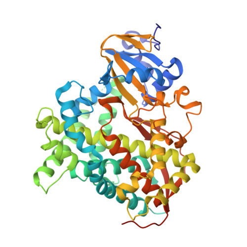
5DYP, 5DYZ - PubMed Abstract:
This paper reports the structure of the double mutant Asp251Gly/Gln307His (named A2) generated by random mutagenesis, able to produce 4'-hydroxydiclofenac, 2-hydroxyibuprofen and 4-hydroxytolbutamide from diclofenac, ibuprofen and tolbutamide, respectively. The 3D structure of the substrate-free mutant shows a conformation similar to the closed one found in the substrate-bound wild type enzyme, but with a higher degree of disorder in the region of the G-helix and F-G loop. This is due to the mutation Asp251Gly that breaks the salt bridge between Aps251 on I-helix and Lys224 on G-helix, allowing the G-helix to move away from I-helix and conferring a higher degree of flexibility to this element. This subtle structural change is accompanied by long-range structural rearrangements of the active site with the rotation of Phe87 and a reorganization of catalytically important water molecules. The impact of these structural features on thermal stability, reduction potential and electron transfer is investigated. The data demonstrate that a single mutation far from the active site triggers an increase in protein flexibility in a key region, shifting the conformational equilibrium toward the closed form that is ready to accept electrons and enter the P450 catalytic cycle as soon as a substrate is accepted.
- Department of Life Sciences and Systems Biology, University of Torino, Via Accademia Albertina 13, Torino, Italy; CrisDi, Interdepartmental Center for Crystallography, University of Torino, Via Pietro Giuria 7, Torino, Italy.
Organizational Affiliation: