5DUS | pdb_00005dus

Crystal structure of MERS-CoV macro domain in complex with ADP-ribose


Experimental Data Snapshot

  • Method: X-RAY DIFFRACTION
  • Resolution: 1.43 Å
  • R-Value Free: 
    0.162 (Depositor), 0.163 (DCC) 
  • R-Value Work: 
    0.127 (Depositor), 0.129 (DCC) 
  • R-Value Observed: 
    0.130 (Depositor) 

wwPDB Validation 3D Report Full Report

Validation slider image for 5DUS

Ligand Structure Quality Assessment 


This is version 1.2 of the entry. See complete history

Literature

Macro Domain from Middle East Respiratory Syndrome Coronavirus (MERS-CoV) Is an Efficient ADP-ribose Binding Module: CRYSTAL STRUCTURE AND BIOCHEMICAL STUDIES

Cho, C.-C.Lin, M.-H.Chuang, C.-Y.Hsu, C.-H.

(2016) J Biological Chem 291: 4894-4902

  • DOI: https://doi.org/10.1074/jbc.M115.700542
  • Primary Citation Related Structures: 
    5DUS

  • PubMed Abstract: 

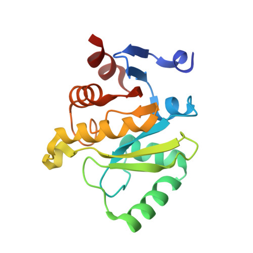
    The newly emerging Middle East respiratory syndrome coronavirus (MERS-CoV) encodes the conserved macro domain within non-structural protein 3. However, the precise biochemical function and structure of the macro domain is unclear. Using differential scanning fluorimetry and isothermal titration calorimetry, we characterized the MERS-CoV macro domain as a more efficient adenosine diphosphate (ADP)-ribose binding module than macro domains from other CoVs. Furthermore, the crystal structure of the MERS-CoV macro domain was determined at 1.43-Å resolution in complex with ADP-ribose. Comparison of macro domains from MERS-CoV and other human CoVs revealed structural differences in the α1 helix alters how the conserved Asp-20 interacts with ADP-ribose and may explain the efficient binding of the MERS-CoV macro domain to ADP-ribose. This study provides structural and biophysical bases to further evaluate the role of the MERS-CoV macro domain in the host response via ADP-ribose binding but also as a potential target for drug design.


  • Organizational Affiliation
    • From the Genome and Systems Biology Degree Program, National Taiwan University and Academia Sinica, Taipei 10617.

Macromolecule Content 

  • Total Structure Weight: 18.81 kDa 
  • Atom Count: 1,492 
  • Modeled Residue Count: 165 
  • Deposited Residue Count: 168 
  • Unique protein chains: 1

Macromolecules

Find similar proteins by:|  3D Structure
Entity ID: 1
MoleculeChains  Sequence LengthOrganismDetailsImage
ORF1a168Middle East respiratory syndrome-related coronavirusMutation(s): 0 
UniProt
Find proteins for K9N638 (Middle East respiratory syndrome-related coronavirus (isolate United Kingdom/H123990006/2012))
Explore K9N638 
Go to UniProtKB:  K9N638
Entity Groups
Sequence Clusters30% Identity50% Identity70% Identity90% Identity95% Identity100% Identity
UniProt GroupK9N638
Sequence Annotations
Expand
Reference Sequence

Experimental Data & Validation

Experimental Data

  • Method: X-RAY DIFFRACTION
  • Resolution: 1.43 Å
  • R-Value Free:  0.162 (Depositor), 0.163 (DCC) 
  • R-Value Work:  0.127 (Depositor), 0.129 (DCC) 
  • R-Value Observed: 0.130 (Depositor) 
Space Group: C 2 2 21
Unit Cell:
Length ( Å )Angle ( ˚ )
a = 41.798α = 90
b = 120.807β = 90
c = 67.659γ = 90
Software Package:
Software NamePurpose
PHENIXrefinement
HKL-2000data reduction
HKL-2000data scaling
PHENIXphasing

Structure Validation

View Full Validation Report


Ligand Structure Quality Assessment 


Entry History 

& Funding Information

Deposition Data


Funding OrganizationLocationGrant Number
the Ministry of Science and TechnologyTaiwan103-2113-M-002-009-MY2
National Taiwan UniversityTaiwanNTU-ERP-104R8600
National Taiwan UniversityTaiwanNTU-ICRP-104R7560-5

Revision History  (Full details and data files)

  • Version 1.0: 2016-01-13
    Type: Initial release
  • Version 1.1: 2016-03-16
    Changes: Database references
  • Version 1.2: 2024-03-20
    Changes: Data collection, Database references, Derived calculations