5DO9

Structure of regulator of G protein signaling 8 (RGS8) in complex with AlF4-activated Galpha-q


Experimental Data Snapshot

  • Method: X-RAY DIFFRACTION
  • Resolution: 2.60 Å
  • R-Value Free: 0.226 
  • R-Value Work: 0.178 
  • R-Value Observed: 0.180 

wwPDB Validation   3D Report Full Report


Ligand Structure Quality Assessment 


This is version 1.5 of the entry. See complete history


Literature

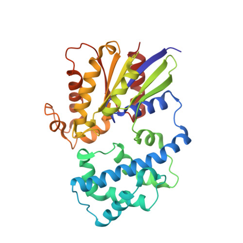
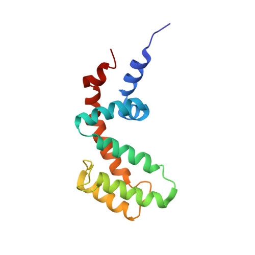
Structure of the Regulator of G Protein Signaling 8 (RGS8)-G alpha q Complex: MOLECULAR BASIS FOR G alpha SELECTIVITY.

Taylor, V.G.Bommarito, P.A.Tesmer, J.J.

(2016) J Biol Chem 291: 5138-5145

  • DOI: https://doi.org/10.1074/jbc.M115.712075
  • Primary Citation of Related Structures:  
    5DO9

  • PubMed Abstract: 

    Regulator of G protein signaling (RGS) proteins interact with activated Gα subunits via their RGS domains and accelerate the hydrolysis of GTP. Although the R4 subfamily of RGS proteins generally accepts both Gαi/o and Gαq/11 subunits as substrates, the R7 and R12 subfamilies select against Gαq/11. In contrast, only one RGS protein, RGS2, is known to be selective for Gαq/11. The molecular basis for this selectivity is not clear. Previously, the crystal structure of RGS2 in complex with Gαq revealed a non-canonical interaction that could be due to interfacial differences imposed by RGS2, the Gα subunit, or both. To resolve this ambiguity, the 2.6 Å crystal structure of RGS8, an R4 subfamily member, was determined in complex with Gαq. RGS8 adopts the same pose on Gαq as it does when bound to Gαi3, indicating that the non-canonical interaction of RGS2 with Gαq is due to unique features of RGS2. Based on the RGS8-Gαq structure, residues in RGS8 that contact a unique α-helical domain loop of Gαq were converted to those typically found in R12 subfamily members, and the reverse substitutions were introduced into RGS10, an R12 subfamily member. Although these substitutions perturbed their ability to stimulate GTP hydrolysis, they did not reverse selectivity. Instead, selectivity for Gαq seems more likely determined by whether strong contacts can be maintained between α6 of the RGS domain and Switch III of Gαq, regions of high sequence and conformational diversity in both protein families.


  • Organizational Affiliation

    From the Life Sciences Institute and the Departments of Pharmacology and Biological Sciences, and the Program in Biophysics, University of Michigan, Ann Arbor, Michigan 48109.


Macromolecules
Find similar proteins by:  (by identity cutoff)  |  3D Structure
Entity ID: 1
MoleculeChains Sequence LengthOrganismDetailsImage
Guanine nucleotide-binding protein G(q) subunit alpha
A, C, E
314Mus musculusMutation(s): 0 
Gene Names: Gnaq
UniProt
Find proteins for P21279 (Mus musculus)
Explore P21279 
Go to UniProtKB:  P21279
Entity Groups  
Sequence Clusters30% Identity50% Identity70% Identity90% Identity95% Identity100% Identity
UniProt GroupP21279
Sequence Annotations
Expand
  • Reference Sequence
Find similar proteins by:  (by identity cutoff)  |  3D Structure
Entity ID: 2
MoleculeChains Sequence LengthOrganismDetailsImage
Regulator of G-protein signaling 8
B, D, F
134Homo sapiensMutation(s): 0 
Gene Names: RGS8
UniProt & NIH Common Fund Data Resources
Find proteins for P57771 (Homo sapiens)
Explore P57771 
Go to UniProtKB:  P57771
PHAROS:  P57771
GTEx:  ENSG00000135824 
Entity Groups  
Sequence Clusters30% Identity50% Identity70% Identity90% Identity95% Identity100% Identity
UniProt GroupP57771
Sequence Annotations
Expand
  • Reference Sequence
Small Molecules
Ligands 3 Unique
IDChains Name / Formula / InChI Key2D Diagram3D Interactions
GDP
Query on GDP

Download Ideal Coordinates CCD File 
G [auth A],
J [auth C],
M [auth E]
GUANOSINE-5'-DIPHOSPHATE
C10 H15 N5 O11 P2
QGWNDRXFNXRZMB-UUOKFMHZSA-N
ALF
Query on ALF

Download Ideal Coordinates CCD File 
H [auth A],
K [auth C],
N [auth E]
TETRAFLUOROALUMINATE ION
Al F4
UYOMQIYKOOHAMK-UHFFFAOYSA-J
MG
Query on MG

Download Ideal Coordinates CCD File 
I [auth A],
L [auth C],
O [auth E]
MAGNESIUM ION
Mg
JLVVSXFLKOJNIY-UHFFFAOYSA-N
Experimental Data & Validation

Experimental Data

  • Method: X-RAY DIFFRACTION
  • Resolution: 2.60 Å
  • R-Value Free: 0.226 
  • R-Value Work: 0.178 
  • R-Value Observed: 0.180 
  • Space Group: C 1 2 1
Unit Cell:
Length ( Å )Angle ( ˚ )
a = 173.946α = 90
b = 95.881β = 94.31
c = 112.898γ = 90
Software Package:
Software NamePurpose
SCALEPACKdata scaling
PHASERphasing
REFMACrefinement
PDB_EXTRACTdata extraction
Cootmodel building
HKL-2000data scaling

Structure Validation

View Full Validation Report


Ligand Structure Quality Assessment 


Entry History 

Deposition Data

Revision History  (Full details and data files)

  • Version 1.0: 2016-01-20
    Type: Initial release
  • Version 1.1: 2016-02-03
    Changes: Database references
  • Version 1.2: 2016-02-10
    Changes: Derived calculations
  • Version 1.3: 2017-11-22
    Changes: Derived calculations, Refinement description
  • Version 1.4: 2018-01-17
    Changes: Author supporting evidence
  • Version 1.5: 2024-03-06
    Changes: Data collection, Database references, Derived calculations, Refinement description