5DLB | pdb_00005dlb

Crystal structure of chaperone EspG3 of ESX-3 type VII secretion system from Mycobacterium marinum M


Experimental Data Snapshot

  • Method: X-RAY DIFFRACTION
  • Resolution: 1.77 Å
  • R-Value Free: 
    0.245 (Depositor), 0.258 (DCC) 
  • R-Value Work: 
    0.209 (Depositor), 0.222 (DCC) 
  • R-Value Observed: 
    0.211 (Depositor) 

wwPDB Validation 3D Report Full Report

Validation slider image for 5DLB

Ligand Structure Quality Assessment 


This is version 1.5 of the entry. See complete history

Literature

Structural Variability of EspG Chaperones from Mycobacterial ESX-1, ESX-3, and ESX-5 Type VII Secretion Systems.

Tuukkanen, A.T.Freire, D.Chan, S.Arbing, M.A.Reed, R.W.Evans, T.J.Zenkeviciute, G.Kim, J.Kahng, S.Sawaya, M.R.Chaton, C.T.Wilmanns, M.Eisenberg, D.Parret, A.H.A.Korotkov, K.V.

(2019) J Mol Biology 431: 289-307

  • DOI: https://doi.org/10.1016/j.jmb.2018.11.003
  • Primary Citation Related Structures: 
    4L4W, 4RCL, 5DLB, 5SXL, 5VBA

  • PubMed Abstract: 

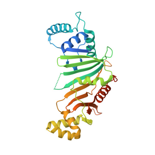
    Type VII secretion systems (ESX) are responsible for transport of multiple proteins in mycobacteria. How different ESX systems achieve specific secretion of cognate substrates remains elusive. In the ESX systems, the cytoplasmic chaperone EspG forms complexes with heterodimeric PE-PPE substrates that are secreted from the cells or remain associated with the cell surface. Here we report the crystal structure of the EspG 1 chaperone from the ESX-1 system determined using a fusion strategy with T4 lysozyme. EspG 1 adopts a quasi 2-fold symmetric structure that consists of a central β-sheet and two α-helical bundles. In addition, we describe the structures of EspG 3 chaperones from four different crystal forms. Alternate conformations of the putative PE-PPE binding site are revealed by comparison of the available EspG 3 structures. Analysis of EspG 1 , EspG 3 , and EspG 5 chaperones using small-angle X-ray scattering reveals that EspG 1 and EspG 3 chaperones form dimers in solution, which we observed in several of our crystal forms. Finally, we propose a model of the ESX-3 specific EspG 3 -PE5-PPE4 complex based on the small-angle X-ray scattering analysis.


  • Organizational Affiliation
    • European Molecular Biology Laboratory, Hamburg Unit, Hamburg 22607, Germany.

Macromolecule Content 

  • Total Structure Weight: 37.94 kDa 
  • Atom Count: 2,149 
  • Modeled Residue Count: 263 
  • Deposited Residue Count: 315 
  • Unique protein chains: 1

Macromolecules

Find similar proteins by:|  3D Structure
Entity ID: 1
MoleculeChains  Sequence LengthOrganismDetailsImage
chaperone EspG3315Mycobacterium marinum MMutation(s): 0 
Gene Names: MMAR_0548
UniProt
Find proteins for B2HNX0 (Mycobacterium marinum (strain ATCC BAA-535 / M))
Explore B2HNX0 
Go to UniProtKB:  B2HNX0
Entity Groups
Sequence Clusters30% Identity50% Identity70% Identity90% Identity95% Identity100% Identity
UniProt GroupB2HNX0
Sequence Annotations
Expand
Reference Sequence

Small Molecules

Ligands 5 Unique
IDChains Name / Formula / InChI Key2D Diagram3D Interactions
CXS

Query on CXS


Download:Ideal Coordinates CCD File
B [auth A]3-CYCLOHEXYL-1-PROPYLSULFONIC ACID
C9 H19 N O3 S
PJWWRFATQTVXHA-UHFFFAOYSA-N
PT

Query on PT


Download:Ideal Coordinates CCD File
DA [auth A]
G [auth A]
IA [auth A]
N [auth A]
NA [auth A]
DA [auth A],
G [auth A],
IA [auth A],
N [auth A],
NA [auth A],
U [auth A],
X [auth A]
PLATINUM (II) ION
Pt
HRGDZIGMBDGFTC-UHFFFAOYSA-N
SO4

Query on SO4


Download:Ideal Coordinates CCD File
E [auth A]SULFATE ION
O4 S
QAOWNCQODCNURD-UHFFFAOYSA-L
GOL

Query on GOL


Download:Ideal Coordinates CCD File
C [auth A],
D [auth A],
F [auth A]
GLYCEROL
C3 H8 O3
PEDCQBHIVMGVHV-UHFFFAOYSA-N
SCN

Query on SCN


Download:Ideal Coordinates CCD File
AA [auth A]
BA [auth A]
CA [auth A]
EA [auth A]
FA [auth A]
AA [auth A],
BA [auth A],
CA [auth A],
EA [auth A],
FA [auth A],
GA [auth A],
H [auth A],
HA [auth A],
I [auth A],
J [auth A],
JA [auth A],
K [auth A],
KA [auth A],
L [auth A],
LA [auth A],
M [auth A],
MA [auth A],
O [auth A],
OA [auth A],
P [auth A],
PA [auth A],
Q [auth A],
QA [auth A],
R [auth A],
RA [auth A],
S [auth A],
SA [auth A],
T [auth A],
V [auth A],
W [auth A],
Y [auth A],
Z [auth A]
THIOCYANATE ION
C N S
ZMZDMBWJUHKJPS-UHFFFAOYSA-M

Experimental Data & Validation

Experimental Data

  • Method: X-RAY DIFFRACTION
  • Resolution: 1.77 Å
  • R-Value Free:  0.245 (Depositor), 0.258 (DCC) 
  • R-Value Work:  0.209 (Depositor), 0.222 (DCC) 
  • R-Value Observed: 0.211 (Depositor) 
Space Group: C 1 2 1
Unit Cell:
Length ( Å )Angle ( ˚ )
a = 112.206α = 90
b = 46.019β = 92.24
c = 58.006γ = 90
Software Package:
Software NamePurpose
REFMACrefinement
XDSdata reduction
XSCALEdata scaling
SHELXphasing

Structure Validation

View Full Validation Report


Ligand Structure Quality Assessment 


Entry History 

Deposition Data

Revision History  (Full details and data files)

  • Version 1.0: 2015-09-23
    Type: Initial release
  • Version 1.1: 2015-09-30
    Changes: Structure summary
  • Version 1.2: 2017-11-01
    Changes: Author supporting evidence, Derived calculations
  • Version 1.3: 2018-12-05
    Changes: Data collection, Database references
  • Version 1.4: 2019-03-06
    Changes: Data collection, Database references
  • Version 1.5: 2024-03-06
    Changes: Data collection, Database references, Derived calculations