


Crystal structure of O-acetylserine sulfhydrylase from haemophilus influenzae in complex with reaction intermediate alpha-aminoacrylate
Kaushik, A., Ekka, M.K., Singh, A.K., Kumaran, S.To be published.
Experimental Data Snapshot
Starting Model: experimental
View more details
Entity ID: 1 | |||||
|---|---|---|---|---|---|
| Molecule | Chains | Sequence Length | Organism | Details | Image |
| Cysteine synthase | A [auth X] | 322 | Haemophilus influenzae Rd KW20 | Mutation(s): 0 Gene Names: cysK, HI_1103 EC: 2.5.1.47 | 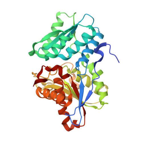 |
UniProt | |||||
Entity Groups | |||||
| Sequence Clusters | 30% Identity50% Identity70% Identity90% Identity95% Identity100% Identity | ||||
| UniProt Group | P45040 | ||||
Sequence AnnotationsExpand | |||||
Reference Sequence | |||||
| Ligands 2 Unique | |||||
|---|---|---|---|---|---|
| ID | Chains | Name / Formula / InChI Key | 2D Diagram | 3D Interactions | |
| 0JO Download:Ideal Coordinates CCD File | C [auth X] | 2-{[(E)-{3-hydroxy-2-methyl-5-[(phosphonooxy)methyl]pyridin-4-yl}methylidene]amino}prop-2-enoic acid C11 H13 N2 O7 P BHIGINKEEFZJGX-YIXHJXPBSA-N |  | ||
| GOL Download:Ideal Coordinates CCD File | B [auth X] | GLYCEROL C3 H8 O3 PEDCQBHIVMGVHV-UHFFFAOYSA-N |  | ||
| Length ( Å ) | Angle ( ˚ ) |
|---|---|
| a = 112.66 | α = 90 |
| b = 112.66 | β = 90 |
| c = 43.369 | γ = 90 |
| Software Name | Purpose |
|---|---|
| PHENIX | refinement |
| HKL-2000 | data reduction |
| HKL-2000 | data scaling |
| PHASER | phasing |


