Characterization of novel small-molecule NRF2 activators: Structural and biochemical validation of stereospecific KEAP1 binding.
Huerta, C., Jiang, X., Trevino, I., Bender, C.F., Ferguson, D.A., Probst, B., Swinger, K.K., Stoll, V.S., Thomas, P.J., Dulubova, I., Visnick, M., Wigley, W.C.(2016) Biochim Biophys Acta 1860: 2537-2552
- PubMed: 27474998 Search on PubMed
- DOI: https://doi.org/10.1016/j.bbagen.2016.07.026
- Primary Citation Related Structures:
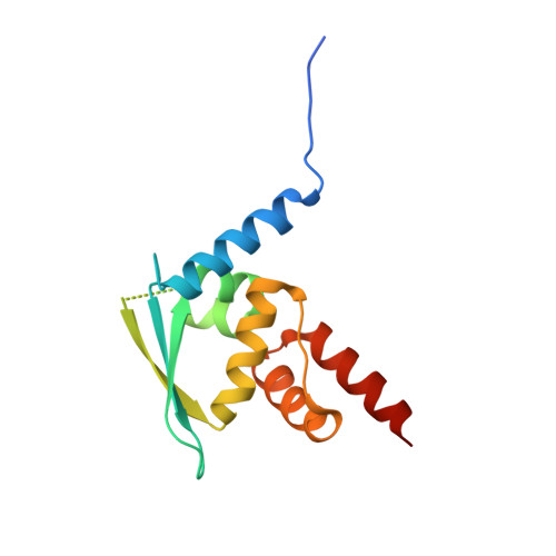
5DAD, 5DAF - PubMed Abstract:
Semi-synthetic oleanane triterpenoid antioxidant inflammation modulators (tpAIMs) are small molecules that interact with KEAP1 cysteine residue 151 (C151) and activate NRF2. Exploration of the structure-activity relationship between the tpAIMs and KEAP1 is limited by the predominantly hydrocarbon nature of the oleanane triterpenoid pentacyclic ring structure. Therefore, we used novel, chemically-tractable, synthetic antioxidant inflammation modulators (sAIMs) to probe the stereoselectivity of the ligand-protein interaction. We measured several parameters of NRF2 activation to assess the potency of sAIM enantiomers with natural (tpAIM-like) 4(S),5(S),10(R) or unnatural 4(R),5(R),10(S) configurations. Additionally, we determined the crystal structure of the KEAP1 BTB domain in complex with two different sAIMs. We found that the potencies of sAIM enantiomers in the natural configuration were similar to those of the tpAIM, RTA 405. Strikingly, sAIM enantiomers in the unnatural configuration were 10- to 40-fold less potent than their natural counterparts. Crystallographic studies of sAIMs in complex with the KEAP1 BTB domain demonstrated that these ligands form a covalent bond with C151 and revealed the presence of additional hydrogen bonds, Van der Waals interactions, and pi-stacking interactions. Although KEAP1 C151 is required for NRF2 activation by tpAIMs and sAIMs, interactions with other KEAP1 residues are critical for the stereospecific recognition and potency of these ligands. This work demonstrates that reversible cyanoenone Michael acceptors, such as the tpAIMs and sAIMs, can be specifically tuned to regulate redox sensitive cysteine residues on key signaling molecules, an approach with significant promise for innovative drug development.
- Department of Research, Reata Pharmaceuticals, Inc., Irving, TX 75063, United States; Department of Physiology, UT Southwestern Medical Center, Dallas, TX 75390, United States.
Organizational Affiliation: