5D7H | pdb_00005d7h

X-RAY CRYSTAL STRUCTURE OF L,D TRANSPEPTIDASE 2 FROM MYCOBACTERIUM TUBERCULOSIS


Experimental Data Snapshot

  • Method: X-RAY DIFFRACTION
  • Resolution: 2.49 Å
  • R-Value Free: 
    0.218 (Depositor), 0.226 (DCC) 
  • R-Value Work: 
    0.162 (Depositor), 0.175 (DCC) 
  • R-Value Observed: 
    0.165 (Depositor) 

wwPDB Validation 3D Report Full Report

Validation slider image for 5D7H

This is version 1.3 of the entry. See complete history

Literature

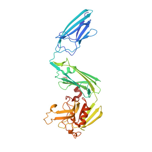
Structural insight into the inactivation of Mycobacterium tuberculosis non-classical transpeptidase LdtMt2 by biapenem and tebipenem.

Bianchet, M.A.Pan, Y.H.Basta, L.A.B.Saavedra, H.Lloyd, E.P.Kumar, P.Mattoo, R.Townsend, C.A.Lamichhane, G.

(2017) BMC Biochem 18: 8-8

  • DOI: https://doi.org/10.1186/s12858-017-0082-4
  • Primary Citation Related Structures: 
    5D7H, 5DC2, 5DCC

  • PubMed Abstract: 

    The carbapenem subclass of β-lactams is among the most potent antibiotics available today. Emerging evidence shows that, unlike other subclasses of β-lactams, carbapenems bind to and inhibit non-classical transpeptidases (L,D-transpeptidases) that generate 3 → 3 linkages in bacterial peptidoglycan. The carbapenems biapenem and tebipenem exhibit therapeutically valuable potencies against Mycobacterium tuberculosis (Mtb). Here, we report the X-ray crystal structures of Mtb L,D-transpeptidase-2 (Ldt Mt2 ) complexed with biapenem or tebipenem. Despite significant variations in carbapenem sulfur side chains, biapenem and tebipenem ultimately form an identical adduct that docks to the outer cavity of Ldt Mt2 . We propose that this common adduct is an enzyme catalyzed decomposition of the carbapenem adduct by a mechanism similar to S-conjugate elimination by β-lyases. The results presented here demonstrate biapenem and tebipenem bind to the outer cavity of Ldt Mt2 , covalently inactivate the enzyme, and subsequently degrade via an S-conjugate elimination mechanism. We discuss structure based drug design based on the findings and propose that the S-conjugate elimination can be leveraged to design novel agents to deliver and locally release antimicrobial factors to act synergistically with the carbapenem carrier.


  • Organizational Affiliation
    • Department of Neurology, Johns Hopkins University School of Medicine, 725 N. Wolfe Street, Baltimore, MD, 21205, USA. bianchet@jhmi.edu.

Macromolecule Content 

  • Total Structure Weight: 77.18 kDa 
  • Atom Count: 5,636 
  • Modeled Residue Count: 702 
  • Deposited Residue Count: 704 
  • Unique protein chains: 1

Macromolecules

Find similar proteins by:|  3D Structure
Entity ID: 1
MoleculeChains  Sequence LengthOrganismDetailsImage
L,D-transpeptidase 2
A, B
352Mycobacterium tuberculosis CDC1551Mutation(s): 0 
Gene Names: ldtBMT2594V735_02606
EC: 2.3.2
UniProt
Find proteins for I6Y9J2 (Mycobacterium tuberculosis (strain ATCC 25618 / H37Rv))
Explore I6Y9J2 
Go to UniProtKB:  I6Y9J2
Entity Groups
Sequence Clusters30% Identity50% Identity70% Identity90% Identity95% Identity100% Identity
UniProt GroupI6Y9J2
Sequence Annotations
Expand
Reference Sequence

Small Molecules

Ligands 2 Unique
IDChains Name / Formula / InChI Key2D Diagram3D Interactions
SO4

Query on SO4


Download:Ideal Coordinates CCD File
C [auth A]
D [auth A]
E [auth A]
F [auth A]
G [auth A]
C [auth A],
D [auth A],
E [auth A],
F [auth A],
G [auth A],
K [auth A],
L [auth B],
M [auth B],
N [auth B],
O [auth B],
P [auth B],
Q [auth B],
R [auth B]
SULFATE ION
O4 S
QAOWNCQODCNURD-UHFFFAOYSA-L
GOL

Query on GOL


Download:Ideal Coordinates CCD File
H [auth A],
I [auth A],
J [auth A],
S [auth B],
T [auth B]
GLYCEROL
C3 H8 O3
PEDCQBHIVMGVHV-UHFFFAOYSA-N

Experimental Data & Validation

Experimental Data

  • Method: X-RAY DIFFRACTION
  • Resolution: 2.49 Å
  • R-Value Free:  0.218 (Depositor), 0.226 (DCC) 
  • R-Value Work:  0.162 (Depositor), 0.175 (DCC) 
  • R-Value Observed: 0.165 (Depositor) 
Space Group: P 1 21 1
Unit Cell:
Length ( Å )Angle ( ˚ )
a = 61.265α = 90
b = 95.579β = 92.59
c = 75.55γ = 90
Software Package:
Software NamePurpose
PHENIXrefinement
PHENIXmodel building
PHENIXphasing

Structure Validation

View Full Validation Report


Entry History 

& Funding Information

Deposition Data


Funding OrganizationLocationGrant Number
National Institutes of Health/National Institute Of Allergy and Infectious Diseases (NIH/NIAID)United StatesR21AI111739
National Institutes of Health/Office of the DirectorUnited StatesDP2OD008459

Revision History  (Full details and data files)

  • Version 1.0: 2016-09-28
    Type: Initial release
  • Version 1.1: 2017-08-23
    Changes: Author supporting evidence, Database references, Derived calculations
  • Version 1.2: 2019-12-11
    Changes: Author supporting evidence
  • Version 1.3: 2024-03-06
    Changes: Data collection, Database references