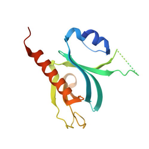
5D3Y

Crystal Structure of the P-Rex1 PH domain with Inositol-(1,3,4,5)-Tetrakisphosphate Bound


Experimental Data Snapshot

  • Method: X-RAY DIFFRACTION
  • Resolution: 1.95 Å
  • R-Value Free: 0.258 
  • R-Value Work: 0.207 
  • R-Value Observed: 0.210 

Starting Model: experimental
View more details

wwPDB Validation   3D Report Full Report


Ligand Structure Quality Assessment 


This is version 1.5 of the entry. See complete history


Literature

Structural and Biochemical Characterization of the Catalytic Core of the Metastatic Factor P-Rex1 and Its Regulation by PtdIns(3,4,5)P3.

Cash, J.N.Davis, E.M.Tesmer, J.J.

(2016) Structure 24: 730-740

  • DOI: https://doi.org/10.1016/j.str.2016.02.022
  • Primary Citation of Related Structures:  
    5D27, 5D3V, 5D3W, 5D3X, 5D3Y, 5FI0, 5FI1

  • PubMed Abstract: 

    Phosphatidylinositol 3,4,5-trisphosphate (PIP3)-dependent Rac exchanger 1 (P-Rex1) is a Rho guanine nucleotide exchange factor synergistically activated by PIP3 and Gβγ that plays an important role in the metastasis of breast, prostate, and skin cancer, making it an attractive therapeutic target. However, the molecular mechanisms behind P-Rex1 regulation are poorly understood. We determined structures of the P-Rex1 pleckstrin homology (PH) domain bound to the headgroup of PIP3 and resolved that PIP3 binding to the PH domain is required for P-Rex1 activity in cells but not for membrane localization, which points to an allosteric activation mechanism by PIP3. We also determined structures of the P-Rex1 tandem Dbl homology/PH domains in complexes with two of its substrate GTPases, Rac1 and Cdc42. Collectively, this study provides important molecular insights into P-Rex1 regulation and tools for targeting the PIP3-binding pocket of P-Rex1 with a new generation of cancer chemotherapeutic agents.


  • Organizational Affiliation

    Departments of Pharmacology and Biological Chemistry, Life Sciences Institute, University of Michigan, Ann Arbor, MI 48109-2216, USA.


Macromolecules
Find similar proteins by:  (by identity cutoff)  |  3D Structure
Entity ID: 1
MoleculeChains Sequence LengthOrganismDetailsImage
Phosphatidylinositol 3,4,5-trisphosphate-dependent Rac exchanger 1 protein
A, B
167Homo sapiensMutation(s): 0 
Gene Names: PREX1KIAA1415
UniProt & NIH Common Fund Data Resources
Find proteins for Q8TCU6 (Homo sapiens)
Explore Q8TCU6 
Go to UniProtKB:  Q8TCU6
PHAROS:  Q8TCU6
GTEx:  ENSG00000124126 
Entity Groups  
Sequence Clusters30% Identity50% Identity70% Identity90% Identity95% Identity100% Identity
UniProt GroupQ8TCU6
Sequence Annotations
Expand
  • Reference Sequence
Experimental Data & Validation

Experimental Data

  • Method: X-RAY DIFFRACTION
  • Resolution: 1.95 Å
  • R-Value Free: 0.258 
  • R-Value Work: 0.207 
  • R-Value Observed: 0.210 
  • Space Group: P 21 21 21
Unit Cell:
Length ( Å )Angle ( ˚ )
a = 59.686α = 90
b = 72.859β = 90
c = 77.939γ = 90
Software Package:
Software NamePurpose
HKL-2000data scaling
HKL-2000data reduction
SCALEPACKdata scaling
PHASERphasing
PHENIXrefinement
Cootmodel building
PDB_EXTRACTdata extraction

Structure Validation

View Full Validation Report


Ligand Structure Quality Assessment 


Entry History & Funding Information

Deposition Data


Funding OrganizationLocationGrant Number
National Institutes of Health/National Heart, Lung, and Blood Institute (NIH/NHLBI)United StatesHL086865
National Institutes of Health/National Heart, Lung, and Blood Institute (NIH/NHLBI)United StatesHL122416
American Cancer SocietyUnited StatesPF-14-224-01-DMC

Revision History  (Full details and data files)

  • Version 1.0: 2016-04-20
    Type: Initial release
  • Version 1.1: 2016-04-27
    Changes: Database references
  • Version 1.2: 2016-05-18
    Changes: Database references
  • Version 1.3: 2017-09-20
    Changes: Author supporting evidence, Database references, Derived calculations, Refinement description
  • Version 1.4: 2019-12-04
    Changes: Author supporting evidence
  • Version 1.5: 2023-09-27
    Changes: Data collection, Database references, Refinement description