5D3E | pdb_00005d3e

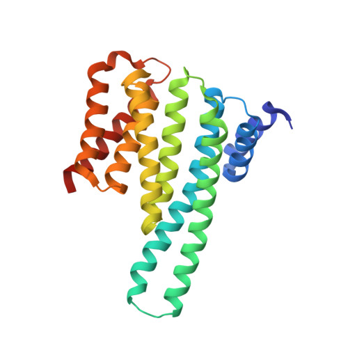
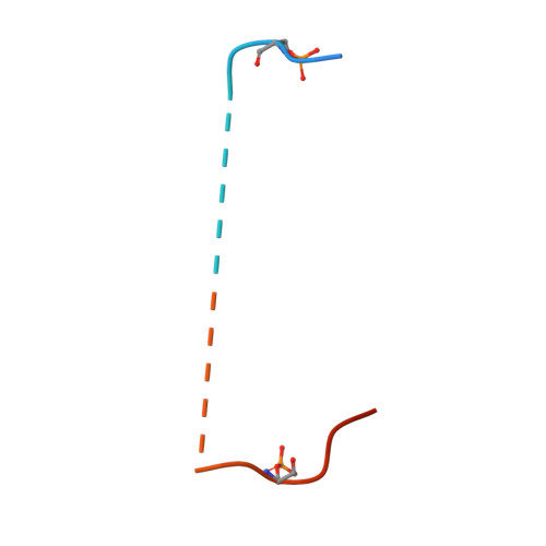
Crystal structure of human 14-3-3 gamma in complex with CFTR R-domain peptide pS768-pS795


Experimental Data Snapshot

  • Method: X-RAY DIFFRACTION
  • Resolution: 2.75 Å
  • R-Value Free: 
    0.244 (Depositor), 0.248 (DCC) 
  • R-Value Work: 
    0.202 (Depositor), 0.206 (DCC) 
  • R-Value Observed: 
    0.204 (Depositor) 

Starting Model: experimental
View more details

wwPDB Validation 3D Report Full Report

Validation slider image for 5D3E

This is version 1.2 of the entry. See complete history. 

Literature

Characterization and small-molecule stabilization of the multisite tandem binding between 14-3-3 and the R domain of CFTR.

Stevers, L.M., Lam, C.V., Leysen, S.F., Meijer, F.A., van Scheppingen, D.S., de Vries, R.M., Carlile, G.W., Milroy, L.G., Thomas, D.Y., Brunsveld, L., Ottmann, C.

(2016) Proc Natl Acad Sci U S A 113: E1152-E1161

  • DOI: https://doi.org/10.1073/pnas.1516631113
  • Primary Citation Related Structures: 
    5D2D, 5D3E, 5D3F

  • PubMed Abstract: 

    Cystic fibrosis is a fatal genetic disease, most frequently caused by the retention of the CFTR (cystic fibrosis transmembrane conductance regulator) mutant protein in the endoplasmic reticulum (ER). The binding of the 14-3-3 protein to the CFTR regulatory (R) domain has been found to enhance CFTR trafficking to the plasma membrane. To define the mechanism of action of this protein-protein interaction, we have examined the interaction in vitro. The disordered multiphosphorylated R domain contains nine different 14-3-3 binding motifs. Furthermore, the 14-3-3 protein forms a dimer containing two amphipathic grooves that can potentially bind these phosphorylated motifs. This results in a number of possible binding mechanisms between these two proteins. Using multiple biochemical assays and crystal structures, we show that the interaction between them is governed by two binding sites: The key binding site of CFTR (pS768) occupies one groove of the 14-3-3 dimer, and a weaker, secondary binding site occupies the other binding groove. We show that fusicoccin-A, a natural-product tool compound used in studies of 14-3-3 biology, can stabilize the interaction between 14-3-3 and CFTR by selectively interacting with a secondary binding motif of CFTR (pS753). The stabilization of this interaction stimulates the trafficking of mutant CFTR to the plasma membrane. This definition of the druggability of the 14-3-3-CFTR interface might offer an approach for cystic fibrosis therapeutics.


  • Organizational Affiliation: 
    • Laboratory of Chemical Biology, Department of Biomedical Engineering, Eindhoven University of Technology, 5600 MB Eindhoven, The Netherlands; Institute for Complex Molecular Systems, Eindhoven University of Technology, 5600 MB Eindhoven, The Netherlands;

Macromolecule Content 

  • Total Structure Weight: 180.43 kDa 
  • Atom Count: 11,713 
  • Modeled Residue Count: 1,431 
  • Deposited Residue Count: 1,566 
  • Unique protein chains: 2

Macromolecules

Find similar proteins by:|  3D Structure
Entity ID: 1
MoleculeChains  Sequence LengthOrganismDetailsImage
14-3-3 protein gamma241Homo sapiensMutation(s): 0 
Gene Names: YWHAG
UniProt & NIH Common Fund Data Resources
Find proteins for P61981 (Homo sapiens)
Explore P61981 
Go to UniProtKB:  P61981
PHAROS:  P61981
GTEx:  ENSG00000170027 
Entity Groups
Sequence Clusters30% Identity50% Identity70% Identity90% Identity95% Identity100% Identity
UniProt GroupP61981
Sequence Annotations
Expand
Reference Sequence
Find similar proteins by:|  3D Structure
Entity ID: 2
MoleculeChains  Sequence LengthOrganismDetailsImage
Cystic fibrosis transmembrane conductance regulatorC,
F [auth G],
I [auth K]
40Homo sapiensMutation(s): 0 
EC: 3.6.3.49 (PDB Primary Data), 5.6.1.6 (UniProt)
UniProt & NIH Common Fund Data Resources
Find proteins for P13569 (Homo sapiens)
Explore P13569 
Go to UniProtKB:  P13569
PHAROS:  P13569
GTEx:  ENSG00000001626 
Entity Groups
Sequence Clusters30% Identity50% Identity70% Identity90% Identity95% Identity100% Identity
UniProt GroupP13569
Sequence Annotations
Expand
Reference Sequence

Small Molecules

Modified Residues  1 Unique
IDChains TypeFormula2D DiagramParent
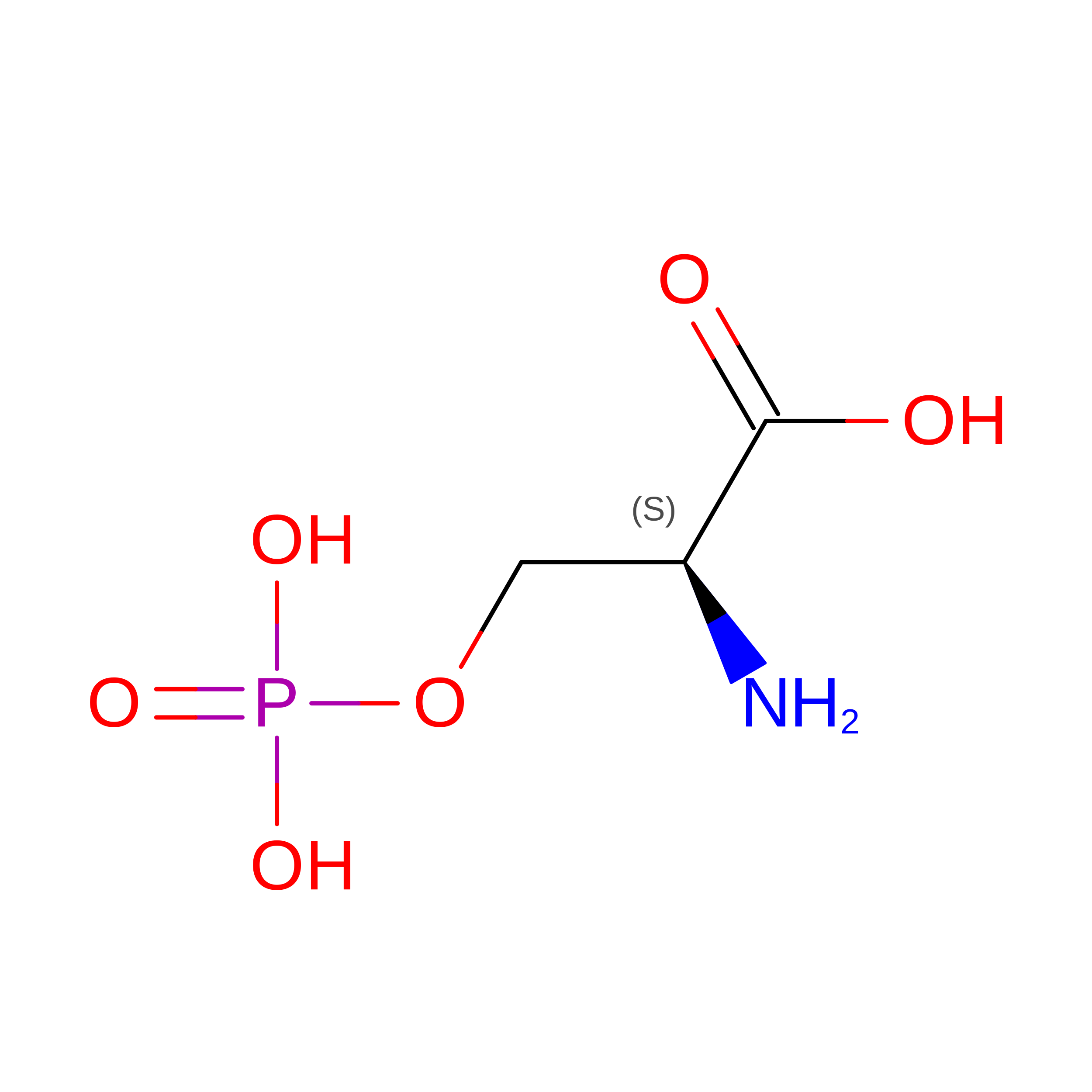
SEP
Query on SEP
C,
F [auth G],
I [auth K]
L-PEPTIDE LINKINGC3 H8 N O6 PSER

Experimental Data & Validation

Experimental Data

  • Method: X-RAY DIFFRACTION
  • Resolution: 2.75 Å
  • R-Value Free:  0.244 (Depositor), 0.248 (DCC) 
  • R-Value Work:  0.202 (Depositor), 0.206 (DCC) 
  • R-Value Observed: 0.204 (Depositor) 
Space Group: P 41 21 2
Unit Cell:
Length ( Å )Angle ( ˚ )
a = 122.45α = 90
b = 122.45β = 90
c = 313.159γ = 90
Software Package:
Software NamePurpose
PHENIXrefinement
XDSdata reduction
Aimlessdata scaling

Structure Validation

View Full Validation Report


Entry History 

Deposition Data

Revision History  (Full details and data files)

  • Version 1.0: 2016-03-16
    Type: Initial release
  • Version 1.1: 2024-01-10
    Changes: Data collection, Database references, Refinement description
  • Version 1.2: 2024-10-16
    Changes: Structure summary