5CZC | pdb_00005czc

The structure of VinK


Experimental Data Snapshot

  • Method: X-RAY DIFFRACTION
  • Resolution: 1.80 Å
  • R-Value Free: 
    0.216 (Depositor), 0.226 (DCC) 
  • R-Value Work: 
    0.165 (Depositor), 0.176 (DCC) 
  • R-Value Observed: 
    0.168 (Depositor) 

wwPDB Validation 3D Report Full Report

Validation slider image for 5CZC

This is version 1.4 of the entry. See complete history

Literature

Structure-based analysis of the molecular interactions between acyltransferase and acyl carrier protein in vicenistatin biosynthesis.

Miyanaga, A.Iwasawa, S.Shinohara, Y.Kudo, F.Eguchi, T.

(2016) Proc Natl Acad Sci U S A 113: 1802-1807

  • DOI: https://doi.org/10.1073/pnas.1520042113
  • Primary Citation Related Structures: 
    5CZC, 5CZD

  • PubMed Abstract: 

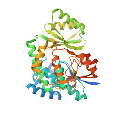
    Acyltransferases (ATs) are key determinants of building block specificity in polyketide biosynthesis. Despite the importance of protein-protein interactions between AT and acyl carrier protein (ACP) during the acyltransfer reaction, the mechanism of ACP recognition by AT is not understood in detail. Herein, we report the crystal structure of AT VinK, which transfers a dipeptide group between two ACPs, VinL and VinP1LdACP, in vicenistatin biosynthesis. The isolated VinK structure showed a unique substrate-binding pocket for the dipeptide group linked to ACP. To gain greater insight into the mechanism of ACP recognition, we attempted to crystallize the VinK-ACP complexes. Because transient enzyme-ACP complexes are difficult to crystallize, we developed a covalent cross-linking strategy using a bifunctional maleimide reagent to trap the VinK-ACP complexes, allowing the determination of the crystal structure of the VinK-VinL complex. In the complex structure, Arg-153, Met-206, and Arg-299 of VinK interact with the negatively charged helix II region of VinL. The VinK-VinL complex structure allows, to our knowledge, the first visualization of the interaction between AT and ACP and provides detailed mechanistic insights into ACP recognition by AT.


  • Organizational Affiliation
    • Department of Chemistry, Tokyo Institute of Technology, O-okayama, Meguro-ku, Tokyo 152-8551, Japan; eguchi@chem.titech.ac.jp miyanaga.a.aa@m.titech.ac.jp.

Macromolecule Content 

  • Total Structure Weight: 72.78 kDa 
  • Atom Count: 5,101 
  • Modeled Residue Count: 601 
  • Deposited Residue Count: 656 
  • Unique protein chains: 1

Macromolecules

Find similar proteins by:|  3D Structure
Entity ID: 1
MoleculeChains  Sequence LengthOrganismDetailsImage
Malonyl-CoA-[acyl-carrier-protein] transacylase
A, B
328Streptomyces halstediiMutation(s): 0 
Gene Names: vinK
EC: 2.3.1.39
UniProt
Find proteins for Q76KY5 (Streptomyces halstedii)
Explore Q76KY5 
Go to UniProtKB:  Q76KY5
Entity Groups
Sequence Clusters30% Identity50% Identity70% Identity90% Identity95% Identity100% Identity
UniProt GroupQ76KY5
Sequence Annotations
Expand
Reference Sequence

Experimental Data & Validation

Experimental Data

  • Method: X-RAY DIFFRACTION
  • Resolution: 1.80 Å
  • R-Value Free:  0.216 (Depositor), 0.226 (DCC) 
  • R-Value Work:  0.165 (Depositor), 0.176 (DCC) 
  • R-Value Observed: 0.168 (Depositor) 
Space Group: P 1 21 1
Unit Cell:
Length ( Å )Angle ( ˚ )
a = 45.52α = 90
b = 74.81β = 91.24
c = 84.81γ = 90
Software Package:
Software NamePurpose
REFMACrefinement
MOSFLMdata reduction
SCALAdata scaling
PHENIXphasing
Cootmodel building

Structure Validation

View Full Validation Report


Entry History 

& Funding Information

Deposition Data


Funding OrganizationLocationGrant Number
Japan Society for the Promotion of ScienceJapan15K18679

Revision History  (Full details and data files)

  • Version 1.0: 2016-02-03
    Type: Initial release
  • Version 1.1: 2016-02-24
    Changes: Database references
  • Version 1.2: 2016-03-30
    Changes: Database references
  • Version 1.3: 2020-02-19
    Changes: Data collection, Database references, Derived calculations
  • Version 1.4: 2024-03-20
    Changes: Data collection, Database references, Derived calculations