5CSE | pdb_00005cse

Streptavidin-S112Y-K121E Complexed with Palladium-Containing Biotin Ligand


Experimental Data Snapshot

  • Method: X-RAY DIFFRACTION
  • Resolution: 1.79 Å
  • R-Value Free: 
    0.256 (Depositor), 0.260 (DCC) 
  • R-Value Work: 
    0.223 (Depositor), 0.227 (DCC) 
  • R-Value Observed: 
    0.225 (Depositor) 

Starting Model: experimental
View more details

wwPDB Validation 3D Report Full Report

Validation slider image for 5CSE

Ligand Structure Quality Assessment 


This is version 2.1 of the entry. See complete history

Literature

An enantioselective artificial Suzukiase based on the biotin-streptavidin technology.

Chatterjee, A.Mallin, H.Klehr, J.Vallapurackal, J.Finke, A.D.Vera, L.Marsh, M.Ward, T.R.

(2016) Chem Sci 7: 673-677

  • DOI: https://doi.org/10.1039/c5sc03116h
  • Primary Citation Related Structures: 
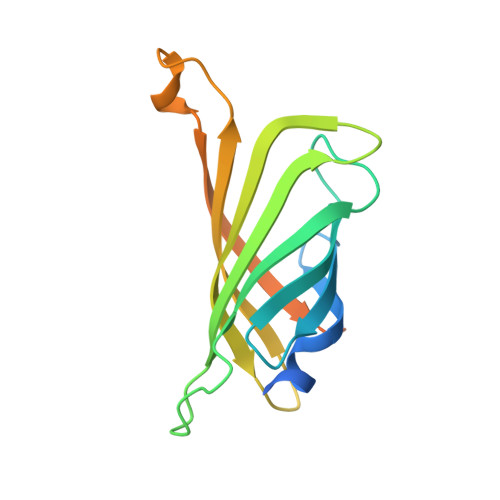
    5CSE

  • PubMed Abstract: 

    Introduction of a biotinylated monophosphine palladium complex within streptavidin affords an enantioselective artificial Suzukiase. Site-directed mutagenesis allowed the optimization of the activity and the enantioselectivity of this artificial metalloenzyme. A variety of atropisomeric biaryls were produced in good yields and up to 90% ee. The hybrid catalyst described herein shows comparable TOF to the previous aqueous-asymmetric Suzuki catalysts, and excellent stability under the reaction conditions to realize higher TON through longer reaction time.


  • Organizational Affiliation
    • Department of Chemistry , University of Basel , Spitalstrasse 51 , 4056 Basel , Switzerland . Email: thomas.ward@unibas.ch.

Macromolecule Content 

  • Total Structure Weight: 34.71 kDa 
  • Atom Count: 1,973 
  • Modeled Residue Count: 243 
  • Deposited Residue Count: 318 
  • Unique protein chains: 1

Macromolecules

Find similar proteins by:|  3D Structure
Entity ID: 1
MoleculeChains  Sequence LengthOrganismDetailsImage
Streptavidin
A, B
159Streptomyces avidiniiMutation(s): 2 
UniProt
Find proteins for P22629 (Streptomyces avidinii)
Explore P22629 
Go to UniProtKB:  P22629
Entity Groups
Sequence Clusters30% Identity50% Identity70% Identity90% Identity95% Identity100% Identity
UniProt GroupP22629
Sequence Annotations
Expand
Reference Sequence

Small Molecules

Ligands 2 Unique
IDChains Name / Formula / InChI Key2D Diagram3D Interactions
SVP

Query on SVP


Download:Ideal Coordinates CCD File
C [auth A],
E [auth B]
chloro{di-tert-butyl[2-({5-[(3aS,4S,6aR)-2-oxohexahydro-1H-thieno[3,4-d]imidazol-4-yl]pentanoyl}amino)ethyl]-lambda~5~-phosphanyl}(1-phenylprop-1-ene-1,3-diyl-kappa~2~C~1~,C~3~)palladium
C29 H46 Cl N3 O2 P Pd S
VFIKYJJBNYQPLX-CRZSKOOBSA-N
CL

Query on CL


Download:Ideal Coordinates CCD File
D [auth A],
F [auth B]
CHLORIDE ION
Cl
VEXZGXHMUGYJMC-UHFFFAOYSA-M

Experimental Data & Validation

Experimental Data

  • Method: X-RAY DIFFRACTION
  • Resolution: 1.79 Å
  • R-Value Free:  0.256 (Depositor), 0.260 (DCC) 
  • R-Value Work:  0.223 (Depositor), 0.227 (DCC) 
  • R-Value Observed: 0.225 (Depositor) 
Space Group: C 2 2 21
Unit Cell:
Length ( Å )Angle ( ˚ )
a = 81.368α = 90
b = 81.46β = 90
c = 90.811γ = 90
Software Package:
Software NamePurpose
PHENIXrefinement
XDSdata reduction
PHASERphasing
XDSdata scaling

Structure Validation

View Full Validation Report


Ligand Structure Quality Assessment 


Entry History 

Deposition Data

Revision History  (Full details and data files)

  • Version 1.0: 2015-11-04
    Type: Initial release
  • Version 2.0: 2018-07-11
    Changes: Atomic model, Data collection, Database references
  • Version 2.1: 2024-01-10
    Changes: Data collection, Database references, Refinement description