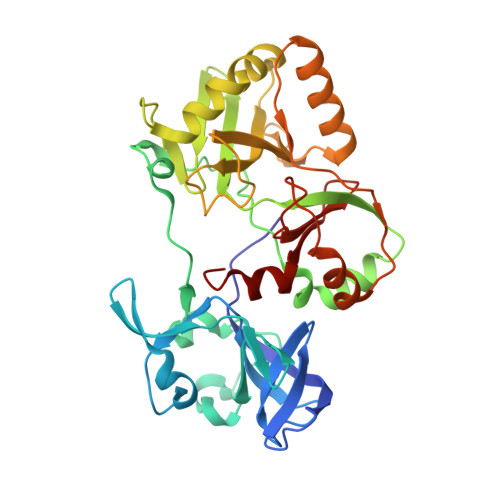
5COT

Structure and mechanism of a eukaryal nick-sealing RNA ligase


Experimental Data Snapshot

  • Method: X-RAY DIFFRACTION
  • Resolution: 1.69 Å
  • R-Value Free: 0.200 
  • R-Value Work: 0.158 
  • R-Value Observed: 0.160 

wwPDB Validation   3D Report Full Report


Ligand Structure Quality Assessment 


This is version 1.4 of the entry. See complete history


Literature

Structure and two-metal mechanism of a eukaryal nick-sealing RNA ligase.

Unciuleac, M.C.Goldgur, Y.Shuman, S.

(2015) Proc Natl Acad Sci U S A 112: 13868-13873

  • DOI: https://doi.org/10.1073/pnas.1516536112
  • Primary Citation of Related Structures:  
    5COT, 5COU, 5COV

  • PubMed Abstract: 

    ATP-dependent RNA ligases are agents of RNA repair that join 3'-OH and 5'-PO4 RNA ends. Naegleria gruberi RNA ligase (NgrRnl) exemplifies a family of RNA nick-sealing enzymes found in bacteria, viruses, and eukarya. Crystal structures of NgrRnl at three discrete steps along the reaction pathway-covalent ligase-(lysyl-Nζ)-AMP•Mn(2+) intermediate; ligase•ATP•(Mn(2+))2 Michaelis complex; and ligase•Mn(2+) complex-highlight a two-metal mechanism of nucleotidyl transfer, whereby (i) an enzyme-bound "catalytic" metal coordination complex lowers the pKa of the lysine nucleophile and stabilizes the transition state of the ATP α phosphate; and (ii) a second metal coordination complex bridges the β- and γ-phosphates. The NgrRnl N domain is a distinctively embellished oligonucleotide-binding (OB) fold that engages the γ-phosphate and associated metal complex and orients the pyrophosphate leaving group for in-line catalysis with stereochemical inversion at the AMP phosphate. The unique domain architecture of NgrRnl fortifies the theme that RNA ligases have evolved many times, and independently, by fusions of a shared nucleotidyltransferase domain to structurally diverse flanking modules. The mechanistic insights to lysine adenylylation gained from the NgrRnl structures are likely to apply broadly to the covalent nucleotidyltransferase superfamily of RNA ligases, DNA ligases, and RNA capping enzymes.


  • Organizational Affiliation

    Molecular Biology Program, Sloan-Kettering Institute, New York, NY 10065;


Macromolecules
Find similar proteins by:  (by identity cutoff)  |  3D Structure
Entity ID: 1
MoleculeChains Sequence LengthOrganismDetailsImage
Naegleria gruberi RNA ligase340Naegleria gruberiMutation(s): 0 
Gene Names: NAEGRDRAFT_82186
UniProt
Find proteins for D2W2Z5 (Naegleria gruberi)
Go to UniProtKB:  D2W2Z5
Sequence Annotations
Expand
  • Reference Sequence
Small Molecules
Ligands 2 Unique
IDChains Name / Formula / InChI Key2D Diagram3D Interactions
AMP
Query on AMP

Download Ideal Coordinates CCD File 
B [auth A]ADENOSINE MONOPHOSPHATE
C10 H14 N5 O7 P
UDMBCSSLTHHNCD-KQYNXXCUSA-N
UNX
Query on UNX

Download Ideal Coordinates CCD File 
C [auth A]UNKNOWN ATOM OR ION
X
Experimental Data & Validation

Experimental Data

  • Method: X-RAY DIFFRACTION
  • Resolution: 1.69 Å
  • R-Value Free: 0.200 
  • R-Value Work: 0.158 
  • R-Value Observed: 0.160 
  • Space Group: P 32
Unit Cell:
Length ( Å )Angle ( ˚ )
a = 55.285α = 90
b = 55.285β = 90
c = 102.545γ = 120
Software Package:
Software NamePurpose
REFMACrefinement
HKL-2000data reduction
HKL-2000data scaling
PHENIXphasing

Structure Validation

View Full Validation Report


Ligand Structure Quality Assessment 


Entry History & Funding Information

Deposition Data


Funding OrganizationLocationGrant Number
National Institutes of Health/National Institute of General Medical Sciences (NIH/NIGMS)United StatesGM42498

Revision History  (Full details and data files)

  • Version 1.0: 2015-10-28
    Type: Initial release
  • Version 1.1: 2015-11-11
    Changes: Database references
  • Version 1.2: 2015-11-25
    Changes: Database references
  • Version 1.3: 2017-09-06
    Changes: Author supporting evidence, Database references, Derived calculations
  • Version 1.4: 2019-12-25
    Changes: Author supporting evidence