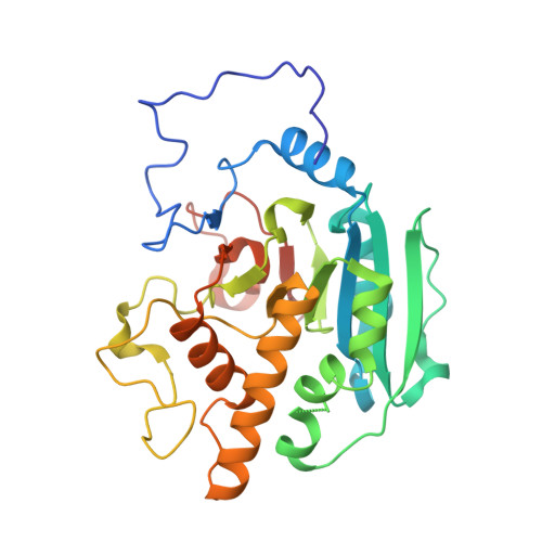
Glycosyltransfer in mutants of putative catalytic residue Glu303 of the human ABO(H) A and B blood group glycosyltransferases GTA and GTB proceeds through a labile active site.
Blackler, R.J., Gagnon, S.M., Polakowski, R., Rose, N.L., Zheng, R.B., Letts, J.A., Johal, A.R., Schuman, B., Borisova, S.N., Palcic, M.M., Evans, S.V.(2017) Glycobiology 27: 370-380
- PubMed: 27979997
- DOI: https://doi.org/10.1093/glycob/cww117
- Primary Citation of Related Structures:
5CMF, 5CMG, 5CMH, 5CMI, 5CMJ, 5CQL, 5CQM, 5CQN, 5CQO, 5CQP - PubMed Abstract:
The homologous glycosyltransferases α-1,3-N-acetylgalactosaminyltransferase (GTA) and α-1,3-galactosyltransferase (GTB) carry out the final synthetic step of the closely related human ABO(H) blood group A and B antigens. The catalytic mechanism of these model retaining enzymes remains under debate, where Glu303 has been suggested to act as a putative nucleophile in a double displacement mechanism, a local dipole stabilizing the intermediate in an orthogonal associative mechanism or a general base to stabilize the reactive oxocarbenium ion-like intermediate in an SNi-like mechanism. Kinetic analysis of GTA and GTB point mutants E303C, E303D, E303Q and E303A shows that despite the enzymes having nearly identical sequences, the corresponding mutants of GTA/GTB have up to a 13-fold difference in their residual activities relative to wild type. High-resolution single crystal X-ray diffraction studies reveal, surprisingly, that the mutated Cys, Asp and Gln functional groups are no more than 0.8 Å further from the anomeric carbon of donor substrate compared to wild type. However, complicating the analysis is the observation that Glu303 itself plays a critical role in maintaining the stability of a strained "double-turn" in the active site through several hydrogen bonds, and any mutation other than E303Q leads to significantly higher thermal motion or even disorder in the substrate recognition pockets. Thus, there is a remarkable juxtaposition of the mutants E303C and E303D, which retain significant activity despite disrupted active site architecture, with GTB/E303Q, which maintains active site architecture but exhibits zero activity. These findings indicate that nucleophilicity at position 303 is more catalytically valuable than active site stability and highlight the mechanistic elasticity of these enzymes.
- Department of Biochemistry and Microbiology, University of Victoria, PO Box 3800, STN CSC, Victoria, BC, Canada.
Organizational Affiliation:


