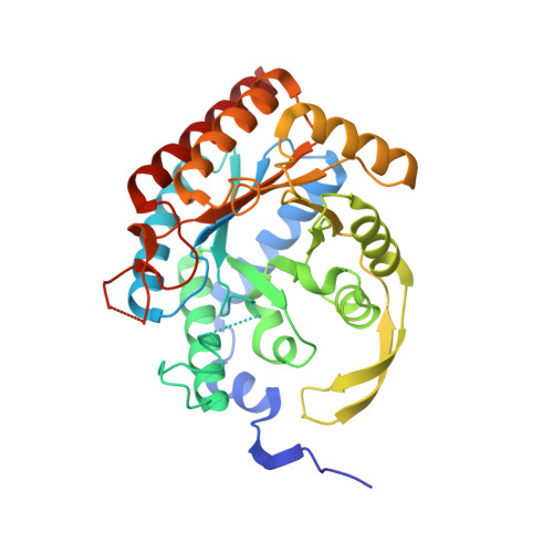
5CKS

DAHP (3-deoxy-D-arabinoheptulosonate-7-phosphate) Synthase in complex with DAHP Oxime.


Experimental Data Snapshot

  • Method: X-RAY DIFFRACTION
  • Resolution: 2.12 Å
  • R-Value Free: 0.238 
  • R-Value Work: 0.181 
  • R-Value Observed: 0.182 

Starting Model: experimental
View more details

wwPDB Validation   3D Report Full Report


Ligand Structure Quality Assessment 

Created with Raphaël 2.3.0Worse 01 BetterLigand structure goodness of fit to experimental dataBest fitted 52LClick on this verticalbar to view detailsBest fitted GALClick on this verticalbar to view details

This is version 1.5 of the entry. See complete history


Literature

Potent Inhibition of 3-Deoxy-d-arabinoheptulosonate-7-phosphate (DAHP) Synthase by DAHP Oxime, a Phosphate Group Mimic.

Balachandran, N.Heimhalt, M.Liuni, P.To, F.Wilson, D.J.Junop, M.S.Berti, P.J.

(2016) Biochemistry 55: 6617-6629

  • DOI: https://doi.org/10.1021/acs.biochem.6b00930
  • Primary Citation of Related Structures:  
    5CKS

  • PubMed Abstract: 

    3-Deoxy-d-arabinoheptulosonate-7-phosphate (DAHP) synthase catalyzes the first step in the shikimate pathway. It catalyzes an aldol-like reaction of phosphoenolpyruvate (PEP) with erythrose 4-phosphate (E4P) to form DAHP. The kinetic mechanism was rapid equilibrium sequential ordered ter ter, with the essential divalent metal ion, Mn 2+ , binding first, followed by PEP and E4P. DAHP oxime, in which an oxime group replaces the keto oxygen, was a potent inhibitor, with K i = 1.5 ± 0.4 μM, though with residual activity at high inhibitor concentrations. It displayed slow-binding inhibition with a residence time, t R , of 83 min. The crystal structure revealed that the oxime functional group, combined with two crystallographic waters, bound at the same location in the catalytic center as the phosphate group of the tetrahedral intermediate. DAHP synthase has a dimer-of-dimers homotetrameric structure, and DAHP oxime bound to only one subunit of each tight dimer. Inhibitor binding was competitive with respect to all three substrates in the subunits to which it bound. DAHP oxime did not overlap with the metal binding site, so the cause of their mutually exclusive binding was not clear. Similarly, there was no obvious structural reason for inhibitor binding in only two subunits; however, changes in global hydrogen/deuterium exchange showed large scale changes in protein dynamics upon inhibitor binding. The k cat value for the residual activity at high inhibitor concentrations was 3-fold lower, and the apparent K M,E4P value decreased at least 10-fold. This positive cooperativity of binding between DAHP oxime in subunits B and C, and E4P in subunits A and D appears to be the dominant cause for incomplete inhibition at high inhibitor concentrations. In spite of its lack of obvious structural similarity to phosphate, the oxime and crystallographic waters acted as a small, neutral phosphate mimic.


  • Organizational Affiliation

    Department of Chemistry, York University , Toronto, Ontario M3J 1P3, Canada.


Macromolecules
Find similar proteins by:  (by identity cutoff)  |  3D Structure
Entity ID: 1
MoleculeChains Sequence LengthOrganismDetailsImage
Phospho-2-dehydro-3-deoxyheptonate aldolase, Phe-sensitive
A, B, C, D
351Escherichia coli K-12Mutation(s): 0 
Gene Names: aroGb0754JW0737
EC: 2.5.1.54
UniProt
Find proteins for P0AB91 (Escherichia coli (strain K12))
Explore P0AB91 
Go to UniProtKB:  P0AB91
Entity Groups  
Sequence Clusters30% Identity50% Identity70% Identity90% Identity95% Identity100% Identity
UniProt GroupP0AB91
Sequence Annotations
Expand
  • Reference Sequence
Experimental Data & Validation

Experimental Data

  • Method: X-RAY DIFFRACTION
  • Resolution: 2.12 Å
  • R-Value Free: 0.238 
  • R-Value Work: 0.181 
  • R-Value Observed: 0.182 
  • Space Group: C 1 2 1
Unit Cell:
Length ( Å )Angle ( ˚ )
a = 209.99α = 90
b = 53.36β = 115.44
c = 150.655γ = 90
Software Package:
Software NamePurpose
PHENIXrefinement
HKL-2000data reduction
HKL-2000data scaling
PHENIXphasing

Structure Validation

View Full Validation Report


Ligand Structure Quality Assessment 

Created with Raphaël 2.3.0Worse 01 BetterLigand structure goodness of fit to experimental dataBest fitted 52LClick on this verticalbar to view detailsBest fitted GALClick on this verticalbar to view details

Entry History & Funding Information

Deposition Data


Funding OrganizationLocationGrant Number
Canadian Institutes of Health Research (CIHR)CanadaMOP-64422
Canadian Institutes of Health Research (CIHR)CanadaMOP-89903

Revision History  (Full details and data files)

  • Version 1.0: 2016-08-10
    Type: Initial release
  • Version 1.1: 2017-01-04
    Changes: Database references
  • Version 1.2: 2017-11-01
    Changes: Author supporting evidence
  • Version 1.3: 2020-01-08
    Changes: Author supporting evidence
  • Version 1.4: 2020-07-29
    Type: Remediation
    Reason: Carbohydrate remediation
    Changes: Data collection, Derived calculations, Structure summary
  • Version 1.5: 2023-09-27
    Changes: Data collection, Database references, Refinement description, Structure summary