5CFU | pdb_00005cfu

Crystal Structure of ANT(2")-Ia in complex with adenylyl-2"-tobramycin


Experimental Data Snapshot

  • Method: X-RAY DIFFRACTION
  • Resolution: 1.82 Å
  • R-Value Free: 
    0.172 (Depositor), 0.184 (DCC) 
  • R-Value Work: 
    0.138 (Depositor), 0.152 (DCC) 
  • R-Value Observed: 
    0.139 (Depositor) 

Starting Model: experimental
View more details

wwPDB Validation 3D Report Full Report

Validation slider image for 5CFU

Ligand Structure Quality Assessment 


This is version 1.4 of the entry. See complete history

Literature

Revisiting the Catalytic Cycle and Kinetic Mechanism of AminoglycosideO-Nucleotidyltransferase(2′′): A Structural and Kinetic Study.

Bassenden, A.V.Park, J.Rodionov, D.Berghuis, A.M.

(2020) ACS Chem Biol 

  • DOI: https://doi.org/10.1021/acschembio.9b00904
  • Primary Citation Related Structures: 
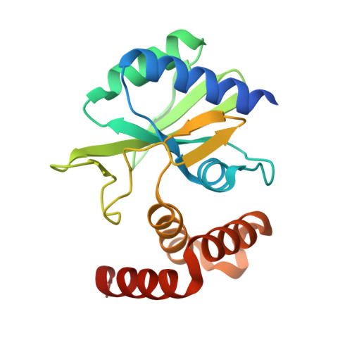
    4XJE, 5CFU

  • PubMed Abstract: 

    Aminoglycoside antibiotics have lost much of their effectiveness due to widespread resistance, primarily via covalent modification. One of the most ubiquitous enzymes responsible for aminoglycoside resistance is aminoglycoside O -nucleotidyltransferase(2″), which catalyzes a nucleotidylation reaction. Due to its clinical importance, much research has focused on dissecting the mechanism of action, some of it dating back more than 30 years. Here, we present structural data for catalytically informative states of the enzyme, i.e., ANT(2″) in complex with adenosine monophosphate (AMP) and tobramycin (inactive-intermediate state) and in complex with adenylyl-2″-tobramycin, pyrophosphate, and Mn 2+ (product-bound state). These two structures in conjunction with our previously reported structure of ANT(2″)'s substrate-bound complex capture clinical states along ANT(2″)'s reaction coordinate. Additionally, isothermal titration calorimetry (ITC)-based studies are presented that assess the order of substrate binding and product release. Combined, these results outline a kinetic mechanism for ANT(2″) that contradicts what has been previously reported. Specifically, we show that the release of adenylated aminoglycoside precedes pyrophosphate. Furthermore, the ternary complex structures provide additional details on the catalytic mechanism, which reveals extensive similarities to the evolutionarily related DNA polymerase-β superfamily.


  • Organizational Affiliation
    • Department of Biochemistry, McGill University, McIntyre Medical Building, 3655 Promenade Sir William Osler, Montréal, Québec Canada, H3G 1Y6.

Macromolecule Content 

  • Total Structure Weight: 22.49 kDa 
  • Atom Count: 1,693 
  • Modeled Residue Count: 174 
  • Deposited Residue Count: 185 
  • Unique protein chains: 1

Macromolecules

Find similar proteins by:|  3D Structure
Entity ID: 1
MoleculeChains  Sequence LengthOrganismDetailsImage
Aminoglycoside Nucleotidyltransferase (2")-Ia185Pseudomonas aeruginosaMutation(s): 0 
Gene Names: aadBTNCP6
UniProt
Find proteins for Q6X3H6 (Pseudomonas aeruginosa)
Explore Q6X3H6 
Go to UniProtKB:  Q6X3H6
Entity Groups
Sequence Clusters30% Identity50% Identity70% Identity90% Identity95% Identity100% Identity
UniProt GroupQ6X3H6
Sequence Annotations
Expand
Reference Sequence

Small Molecules

Ligands 4 Unique
IDChains Name / Formula / InChI Key2D Diagram3D Interactions
51H

Query on 51H


Download:Ideal Coordinates CCD File
D [auth A]adenylyl-2"-tobramycin
C28 H49 N10 O15 P
RHQLHNATZATWNZ-XMUWNKPOSA-N
POP

Query on POP


Download:Ideal Coordinates CCD File
E [auth A],
F [auth A]
PYROPHOSPHATE 2-
H2 O7 P2
XPPKVPWEQAFLFU-UHFFFAOYSA-L
BU1

Query on BU1


Download:Ideal Coordinates CCD File
G [auth A],
H [auth A],
I [auth A]
1,4-BUTANEDIOL
C4 H10 O2
WERYXYBDKMZEQL-UHFFFAOYSA-N
MN

Query on MN


Download:Ideal Coordinates CCD File
B [auth A],
C [auth A]
MANGANESE (II) ION
Mn
WAEMQWOKJMHJLA-UHFFFAOYSA-N

Experimental Data & Validation

Experimental Data

  • Method: X-RAY DIFFRACTION
  • Resolution: 1.82 Å
  • R-Value Free:  0.172 (Depositor), 0.184 (DCC) 
  • R-Value Work:  0.138 (Depositor), 0.152 (DCC) 
  • R-Value Observed: 0.139 (Depositor) 
Space Group: P 1 21 1
Unit Cell:
Length ( Å )Angle ( ˚ )
a = 46.09α = 90
b = 41.91β = 105.08
c = 47.13γ = 90
Software Package:
Software NamePurpose
PHENIXrefinement
StructureStudiodata collection
xia2data scaling
XDSdata reduction
XSCALEdata scaling
PHASERphasing
PDB_EXTRACTdata extraction

Structure Validation

View Full Validation Report


Ligand Structure Quality Assessment 


Entry History 

& Funding Information

Deposition Data


Funding OrganizationLocationGrant Number
Canadian Institutes of Health Research (CIHR)CanadaCIHR MOP-13107

Revision History  (Full details and data files)

  • Version 1.0: 2016-07-20
    Type: Initial release
  • Version 1.1: 2017-11-22
    Changes: Derived calculations, Refinement description
  • Version 1.2: 2020-01-08
    Changes: Author supporting evidence
  • Version 1.3: 2020-03-18
    Changes: Database references
  • Version 1.4: 2023-09-27
    Changes: Data collection, Database references, Refinement description