5CFF

Crystal structure of Miranda/Staufen dsRBD5 complex


Experimental Data Snapshot

  • Method: X-RAY DIFFRACTION
  • Resolution: 2.50 Å
  • R-Value Free: 0.281 
  • R-Value Work: 0.242 
  • R-Value Observed: 0.244 

wwPDB Validation   3D Report Full Report


This is version 1.1 of the entry. See complete history


Literature

The structural basis of Miranda-mediated Staufen localization during Drosophila neuroblast asymmetric division

Jia, M.Shan, Z.Yang, Y.Liu, C.Li, J.Luo, Z.G.Zhang, M.Cai, Y.Wen, W.Wang, W.

(2015) Nat Commun 6: 8381-8381

  • DOI: https://doi.org/10.1038/ncomms9381
  • Primary Citation of Related Structures:  
    5CFF

  • PubMed Abstract: 

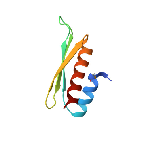
    During the asymmetric division of Drosophila neuroblasts (NBs), the scaffold Miranda (Mira) coordinates the subcellular distribution of cell-fate determinants including Staufen (Stau) and segregates them into the ganglion mother cells (GMCs). Here we show the fifth double-stranded RNA (dsRNA)-binding domain (dsRBD5) of Stau is necessary and sufficient for binding to a coiled-coil region of Mira cargo-binding domain (CBD). The crystal structure of Mira514-595/Stau dsRBD5 complex illustrates that Mira forms an elongated parallel coiled-coil dimer, and two dsRBD5 symmetrically bind to the Mira dimer through their exposed β-sheet faces, revealing a previously unrecognized protein interaction mode for dsRBDs. We further demonstrate that the Mira-Stau dsRBD5 interaction is responsible for the asymmetric localization of Stau during Drosophila NB asymmetric divisions. Finally, we find the CBD-mediated dimer assembly is likely a common requirement for Mira to recognize and translocate other cargos including brain tumour (Brat).


  • Organizational Affiliation

    Shanghai Key Laboratory of Molecular Catalysis and Innovative Materials, Department of Chemistry and Key Laboratory of Molecular Medicine, Ministry of Education, Institutes of Biomedical Sciences, Shanghai Medical College, Fudan University, Shanghai 200433, China.


Macromolecules
Find similar proteins by:  (by identity cutoff)  |  3D Structure
Entity ID: 1
MoleculeChains Sequence LengthOrganismDetailsImage
Miranda
A, B, C, D
95Drosophila melanogasterMutation(s): 0 
Gene Names: miraCG12249Dmel_CG12249
UniProt
Find proteins for Q9VDR7 (Drosophila melanogaster)
Explore Q9VDR7 
Go to UniProtKB:  Q9VDR7
Entity Groups  
Sequence Clusters30% Identity50% Identity70% Identity90% Identity95% Identity100% Identity
UniProt GroupQ9VDR7
Sequence Annotations
Expand
  • Reference Sequence
Find similar proteins by:  (by identity cutoff)  |  3D Structure
Entity ID: 2
MoleculeChains Sequence LengthOrganismDetailsImage
Staufen
E, F, G, H
72Drosophila melanogasterMutation(s): 0 
Gene Names: stauCG5753
UniProt
Find proteins for P25159 (Drosophila melanogaster)
Explore P25159 
Go to UniProtKB:  P25159
Entity Groups  
Sequence Clusters30% Identity50% Identity70% Identity90% Identity95% Identity100% Identity
UniProt GroupP25159
Sequence Annotations
Expand
  • Reference Sequence
Small Molecules
Modified Residues  1 Unique
IDChains TypeFormula2D DiagramParent
MSE
Query on MSE
A, B, C, D
L-PEPTIDE LINKINGC5 H11 N O2 SeMET
Experimental Data & Validation

Experimental Data

  • Method: X-RAY DIFFRACTION
  • Resolution: 2.50 Å
  • R-Value Free: 0.281 
  • R-Value Work: 0.242 
  • R-Value Observed: 0.244 
  • Space Group: C 1 2 1
Unit Cell:
Length ( Å )Angle ( ˚ )
a = 200.023α = 90
b = 51.332β = 90.56
c = 100.274γ = 90
Software Package:
Software NamePurpose
HKL-2000data collection
HKL-2000data scaling
SHELXphasing
DMphasing
PHENIXrefinement
PDB_EXTRACTdata extraction
HKL-2000data reduction

Structure Validation

View Full Validation Report


Entry History 

Deposition Data

Revision History  (Full details and data files)

  • Version 1.0: 2015-10-21
    Type: Initial release
  • Version 1.1: 2015-10-28
    Changes: Database references