5C8O | pdb_00005c8o

Crystal structure of MoCVNH3 variant (Mo0v)


Experimental Data Snapshot

  • Method: X-RAY DIFFRACTION
  • Resolution: 2.09 Å
  • R-Value Free: 
    0.281 (Depositor), 0.284 (DCC) 
  • R-Value Work: 
    0.220 (Depositor), 0.229 (DCC) 
  • R-Value Observed: 
    0.226 (Depositor) 

Starting Models: experimental
View more details

wwPDB Validation 3D Report Full Report

Validation slider image for 5C8O

This is version 1.5 of the entry. See complete history

Literature

Structural Insight into Fungal Cell Wall Recognition by a CVNH Protein with a Single LysM Domain.

Koharudin, L.M.Debiec, K.T.Gronenborn, A.M.

(2015) Structure 23: 2143-2154

  • DOI: https://doi.org/10.1016/j.str.2015.07.023
  • Primary Citation Related Structures: 
    5C8O, 5C8P, 5C8Q

  • PubMed Abstract: 

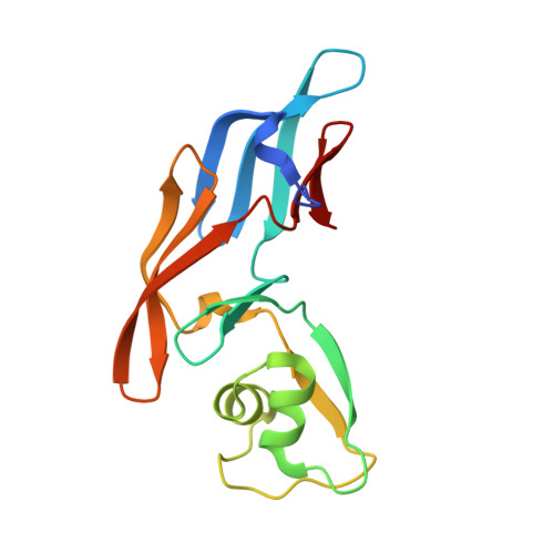
    MGG_03307 is a lectin isolated from Magnaporte oryzae, a fungus that causes devastating rice blast disease. Its function is associated with protecting M. oryzae from the host immune response in plants. To provide the structural basis of how MGG_03307 protects the fungus, crystal structures of its CVNH-LysM module were determined in the absence and presence of GlcNAc-containing cell wall chitin constituents, which can act as pathogen-associated molecular patterns. Our structures revealed that glycan binding is accompanied by a notable conformational change in the LysM domain and that GlcNAc3 and GlcNAc4 are accommodated similarly. GlcNAc5 and GlcNAc6 interact with the LysM domain in multiple conformations, as evidenced by solution nuclear magnetic resonance studies. No dimerization of MoCVNH3 via its LysM domain was observed upon binding to GlcNAc6, unlike in multiple LysM domain-containing proteins. Importantly, we define a specific consensus binding mode for the recognition of GlcNAc oligomers by single LysM domains.


  • Organizational Affiliation
    • Department of Structural Biology, University of Pittsburgh School of Medicine, 3501 Fifth Avenue, Pittsburgh, PA 15260, USA.

Macromolecule Content 

  • Total Structure Weight: 17.35 kDa 
  • Atom Count: 1,329 
  • Modeled Residue Count: 151 
  • Deposited Residue Count: 154 
  • Unique protein chains: 1

Macromolecules

Find similar proteins by:|  3D Structure
Entity ID: 1
MoleculeChains  Sequence LengthOrganismDetailsImage
MoCVNH3 variant154Pyricularia oryzaeMutation(s): 1 
UniProt
Find proteins for G4N991 (Pyricularia oryzae (strain 70-15 / ATCC MYA-4617 / FGSC 8958))
Explore G4N991 
Go to UniProtKB:  G4N991
Entity Groups
Sequence Clusters30% Identity50% Identity70% Identity90% Identity95% Identity100% Identity
UniProt GroupG4N991
Sequence Annotations
Expand
Reference Sequence

Experimental Data & Validation

Experimental Data

  • Method: X-RAY DIFFRACTION
  • Resolution: 2.09 Å
  • R-Value Free:  0.281 (Depositor), 0.284 (DCC) 
  • R-Value Work:  0.220 (Depositor), 0.229 (DCC) 
  • R-Value Observed: 0.226 (Depositor) 
Space Group: P 1 21 1
Unit Cell:
Length ( Å )Angle ( ˚ )
a = 37.411α = 90
b = 51.075β = 110.62
c = 43.243γ = 90
Software Package:
Software NamePurpose
REFMACrefinement
d*TREKdata reduction
Cootmodel building
d*TREKdata scaling
PHASERphasing

Structure Validation

View Full Validation Report


Entry History 

& Funding Information

Deposition Data


Funding OrganizationLocationGrant Number
National Institutes of Health/National Institute of General Medical Sciences (NIH/NIGMS)United StatesGM080642

Revision History  (Full details and data files)

  • Version 1.0: 2015-08-12
    Type: Initial release
  • Version 1.1: 2015-10-28
    Changes: Database references
  • Version 1.2: 2015-11-11
    Changes: Database references
  • Version 1.3: 2017-09-27
    Changes: Author supporting evidence, Database references, Derived calculations
  • Version 1.4: 2019-12-25
    Changes: Author supporting evidence
  • Version 1.5: 2023-09-27
    Changes: Data collection, Database references, Refinement description