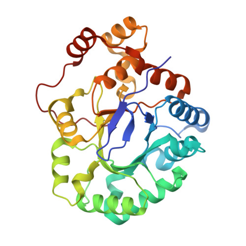
Crystal structure of aldo-keto reductase from Sinorhizobium meliloti 1021 in complex with NADPH
Gasiorowska, O.A., Shabalin, I.G., Handing, K.B., Seidel, R., Bonanno, J., Almo, S.C., Minor, W., New York Structural Genomics Research Consortium (NYSGRC)To be published.