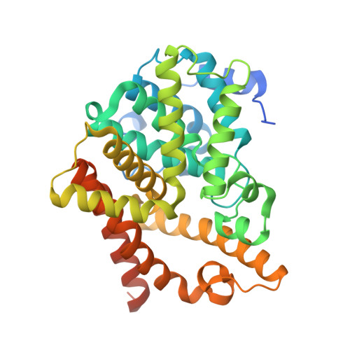
5C2E

PDE10 complexed with6-chloro-N-[(2,4-dimethylthiazol-5-yl)methyl]-5-methyl-2-[2-(2-pyridyl)ethoxy]pyrimidin-4-amine


Experimental Data Snapshot

  • Method: X-RAY DIFFRACTION
  • Resolution: 2.10 Å
  • R-Value Free: 0.280 
  • R-Value Work: 0.249 
  • R-Value Observed: 0.251 

Starting Model: experimental
View more details

wwPDB Validation   3D Report Full Report


Ligand Structure Quality Assessment 


This is version 1.2 of the entry. See complete history


Literature

Discovery and Optimization of a Series of Pyrimidine-Based Phosphodiesterase 10A (PDE10A) Inhibitors through Fragment Screening, Structure-Based Design, and Parallel Synthesis.

Shipe, W.D.Sharik, S.S.Barrow, J.C.McGaughey, G.B.Theberge, C.R.Uslaner, J.M.Yan, Y.Renger, J.J.Smith, S.M.Coleman, P.J.Cox, C.D.

(2015) J Med Chem 58: 7888-7894

  • DOI: https://doi.org/10.1021/acs.jmedchem.5b00983
  • Primary Citation of Related Structures:  
    5C1W, 5C28, 5C29, 5C2A, 5C2E, 5C2H

  • PubMed Abstract: 

    Screening of a fragment library for PDE10A inhibitors identified a low molecular weight pyrimidine hit with PDE10A Ki of 8700 nM and LE of 0.59. Initial optimization by catalog followed by iterative parallel synthesis guided by X-ray cocrystal structures resulted in rapid potency improvements with minimal loss of ligand efficiency. Compound 15 h, with PDE10A Ki of 8.2 pM, LE of 0.49, and >5000-fold selectivity over other PDEs, fully attenuates MK-801-induced hyperlocomotor activity after ip dosing.


  • Organizational Affiliation

    Departments of †Discovery Chemistry, ‡Chemistry Modeling and Informatics, §Neuroscience, and ∥Structural Chemistry, Merck Research Laboratories , West Point, Pennsylvania 19486, United States.


Macromolecules
Find similar proteins by:  (by identity cutoff)  |  3D Structure
Entity ID: 1
MoleculeChains Sequence LengthOrganismDetailsImage
cAMP and cAMP-inhibited cGMP 3',5'-cyclic phosphodiesterase 10A
A, B
362Homo sapiensMutation(s): 0 
Gene Names: PDE10A
EC: 3.1.4.17 (PDB Primary Data), 3.1.4.35 (PDB Primary Data)
UniProt & NIH Common Fund Data Resources
Find proteins for Q9Y233 (Homo sapiens)
Explore Q9Y233 
Go to UniProtKB:  Q9Y233
PHAROS:  Q9Y233
GTEx:  ENSG00000112541 
Entity Groups  
Sequence Clusters30% Identity50% Identity70% Identity90% Identity95% Identity100% Identity
UniProt GroupQ9Y233
Sequence Annotations
Expand
  • Reference Sequence
Small Molecules
Ligands 4 Unique
IDChains Name / Formula / InChI Key2D Diagram3D Interactions
4Y1
Query on 4Y1

Download Ideal Coordinates CCD File 
H [auth B]6-chloro-N-[(2,4-dimethyl-1,3-thiazol-5-yl)methyl]-5-methyl-2-[2-(pyridin-2-yl)ethoxy]pyrimidin-4-amine
C18 H20 Cl N5 O S
SAFPHTULZQIFPO-UHFFFAOYSA-N
4PX
Query on 4PX

Download Ideal Coordinates CCD File 
E [auth A]3-(1-hydroxy-2-methylpropan-2-yl)-5-phenyl-3,5-dihydro-1H-imidazo[4,5-c][1,8]naphthyridine-2,4-dione
C19 H18 N4 O3
ZQRNBXXBVMUYDO-UHFFFAOYSA-N
ZN
Query on ZN

Download Ideal Coordinates CCD File 
C [auth A],
F [auth B]
ZINC ION
Zn
PTFCDOFLOPIGGS-UHFFFAOYSA-N
MG
Query on MG

Download Ideal Coordinates CCD File 
D [auth A],
G [auth B]
MAGNESIUM ION
Mg
JLVVSXFLKOJNIY-UHFFFAOYSA-N
Experimental Data & Validation

Experimental Data

  • Method: X-RAY DIFFRACTION
  • Resolution: 2.10 Å
  • R-Value Free: 0.280 
  • R-Value Work: 0.249 
  • R-Value Observed: 0.251 
  • Space Group: P 21 21 21
Unit Cell:
Length ( Å )Angle ( ˚ )
a = 50.5α = 90
b = 81.35β = 90
c = 158.4γ = 90
Software Package:
Software NamePurpose
BUSTER-TNTrefinement
SCALEPACKdata scaling
PDB_EXTRACTdata extraction
HKL-2000data reduction
REFMACphasing

Structure Validation

View Full Validation Report


Ligand Structure Quality Assessment 


Entry History 

Deposition Data

  • Released Date: 2015-12-02 
  • Deposition Author(s): Yan, Y.

Revision History  (Full details and data files)

  • Version 1.0: 2015-12-02
    Type: Initial release
  • Version 1.1: 2023-09-27
    Changes: Data collection, Database references, Derived calculations, Refinement description
  • Version 1.2: 2024-03-13
    Changes: Source and taxonomy