5BYA | pdb_00005bya

Crystal structure of the catalytic domain of human diphosphoinositol pentakisphosphate kinase 2 (PPIP5K2) in complex with ADP and 1,5-(PCP)2-IP4


Experimental Data Snapshot

  • Method: X-RAY DIFFRACTION
  • Resolution: 1.90 Å
  • R-Value Free: 
    0.199 (Depositor), 0.199 (DCC) 
  • R-Value Work: 
    0.141 (Depositor), 0.142 (DCC) 
  • R-Value Observed: 
    0.145 (Depositor) 

Starting Model: experimental
View more details

wwPDB Validation 3D Report Full Report

Validation slider image for 5BYA

Ligand Structure Quality Assessment 


This is version 1.1 of the entry. See complete history

Literature

Synthetic tools for studying the chemical biology of InsP8.

Riley, A.M.Wang, H.Shears, S.B.L Potter, B.V.

(2015) Chem Commun (Camb) 51: 12605-12608

  • DOI: https://doi.org/10.1039/c5cc05017k
  • Primary Citation Related Structures: 
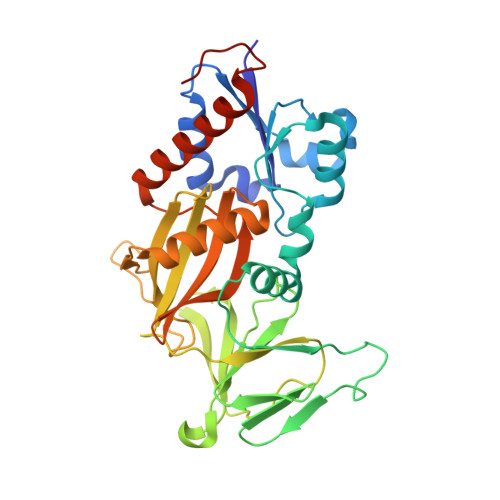
    5BYA, 5BYB

  • PubMed Abstract: 

    To synthesise stabilised mimics of InsP8, the most phosphorylated inositol phosphate signalling molecule in Nature, we replaced its two diphosphate (PP) groups with either phosphonoacetate (PA) or methylenebisphosphonate (PCP) groups. Utility of the PA and PCP analogues was verified by structural and biochemical analyses of their interactions with enzymes of InsP8 metabolism.


  • Organizational Affiliation
    • Wolfson Laboratory of Medicinal Chemistry, Department of Pharmacy and Pharmacology, University of Bath, Claverton Down, Bath, BA2 7AY, UK.

Macromolecule Content 

  • Total Structure Weight: 39.48 kDa 
  • Atom Count: 3,154 
  • Modeled Residue Count: 318 
  • Deposited Residue Count: 330 
  • Unique protein chains: 1

Macromolecules

Find similar proteins by:|  3D Structure
Entity ID: 1
MoleculeChains  Sequence LengthOrganismDetailsImage
Inositol hexakisphosphate and diphosphoinositol-pentakisphosphate kinase 2330Homo sapiensMutation(s): 0 
Gene Names: PPIP5K2HISPPD1KIAA0433VIP2
EC: 2.7.4.21 (PDB Primary Data), 2.7.4.24 (PDB Primary Data)
UniProt & NIH Common Fund Data Resources
Find proteins for O43314 (Homo sapiens)
Explore O43314 
Go to UniProtKB:  O43314
PHAROS:  O43314
GTEx:  ENSG00000145725 
Entity Groups
Sequence Clusters30% Identity50% Identity70% Identity90% Identity95% Identity100% Identity
UniProt GroupO43314
Sequence Annotations
Expand
Reference Sequence

Small Molecules

Ligands 5 Unique
IDChains Name / Formula / InChI Key2D Diagram3D Interactions
4WZ

Query on 4WZ


Download:Ideal Coordinates CCD File
C [auth A]{[(1R,3S,4S,5R,6S)-2,4,5,6-tetrakis(phosphonooxy)cyclohexane-1,3-diyl]bis[oxy(hydroxyphosphoryl)methanediyl]}bis(phosphonic acid)
C8 H24 O28 P8
WDYISGXURTYYJP-ZDJZFTHGSA-N
ADP

Query on ADP


Download:Ideal Coordinates CCD File
B [auth A]ADENOSINE-5'-DIPHOSPHATE
C10 H15 N5 O10 P2
XTWYTFMLZFPYCI-KQYNXXCUSA-N
EDO

Query on EDO


Download:Ideal Coordinates CCD File
I [auth A],
K [auth A],
L [auth A],
M [auth A],
O [auth A]
1,2-ETHANEDIOL
C2 H6 O2
LYCAIKOWRPUZTN-UHFFFAOYSA-N
ACT

Query on ACT


Download:Ideal Coordinates CCD File
J [auth A],
N [auth A],
P [auth A],
Q [auth A]
ACETATE ION
C2 H3 O2
QTBSBXVTEAMEQO-UHFFFAOYSA-M
MG

Query on MG


Download:Ideal Coordinates CCD File
D [auth A],
E [auth A],
F [auth A],
G [auth A],
H [auth A]
MAGNESIUM ION
Mg
JLVVSXFLKOJNIY-UHFFFAOYSA-N

Experimental Data & Validation

Experimental Data

  • Method: X-RAY DIFFRACTION
  • Resolution: 1.90 Å
  • R-Value Free:  0.199 (Depositor), 0.199 (DCC) 
  • R-Value Work:  0.141 (Depositor), 0.142 (DCC) 
  • R-Value Observed: 0.145 (Depositor) 
Space Group: P 21 21 21
Unit Cell:
Length ( Å )Angle ( ˚ )
a = 88.567α = 90
b = 110.4β = 90
c = 41.455γ = 90
Software Package:
Software NamePurpose
HKL-2000data reduction
HKL-2000data scaling
REFMACrefinement
PDB_EXTRACTdata extraction
PHASERphasing

Structure Validation

View Full Validation Report


Ligand Structure Quality Assessment 


Entry History 

Deposition Data

Revision History  (Full details and data files)

  • Version 1.0: 2015-07-22
    Type: Initial release
  • Version 1.1: 2023-09-27
    Changes: Data collection, Database references, Derived calculations, Refinement description