5BUU | pdb_00005buu

Crystal structure of the GluA2 ligand-binding domain (L483Y-N754S) in complex with glutamate and BPAM-321 at 2.07 A resolution


Experimental Data Snapshot

  • Method: X-RAY DIFFRACTION
  • Resolution: 2.07 Å
  • R-Value Free: 
    0.250 (Depositor), 0.205 (DCC) 
  • R-Value Work: 
    0.204 (Depositor), 0.209 (DCC) 
  • R-Value Observed: 
    0.206 (Depositor) 

Starting Model: experimental
View more details

wwPDB Validation 3D Report Full Report

Validation slider image for 5BUU

Ligand Structure Quality Assessment 


This is version 1.4 of the entry. See complete history

Literature

Synthesis and Pharmacology of Mono-, Di-, and Trialkyl-Substituted 7-Chloro-3,4-dihydro-2H-1,2,4-benzothiadiazine 1,1-Dioxides Combined with X-ray Structure Analysis to Understand the Unexpected Structure-Activity Relationship at AMPA Receptors.

Larsen, A.P.Francotte, P.Frydenvang, K.Tapken, D.Goffin, E.Fraikin, P.Caignard, D.H.Lestage, P.Danober, L.Pirotte, B.Kastrup, J.S.

(2016) ACS Chem Neurosci 7: 378-390

  • DOI: https://doi.org/10.1021/acschemneuro.5b00318
  • Primary Citation Related Structures: 
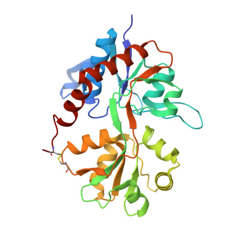
    5BUU

  • PubMed Abstract: 

    Positive allosteric modulators of 2-amino-3-(3-hydroxy-5-methylisoxazol-4-yl)propionic acid (AMPA)-type ionotropic glutamate receptors are promising compounds for treatment of neurological disorders, for example, Alzheimer's disease. Here, we report synthesis and pharmacological evaluation of a series of mono-, di-, or trialkyl-substituted 7-chloro-3,4-dihydro-2H-1,2,4-benzothiadiazine 1,1-dioxides, comprising in total 16 new modulators. The trisubstituted compounds 7b, 7d, and 7e revealed potent activity (EC2× = 2.7-4.3 μM; concentration of compound responsible for a 2-fold increase of the AMPA mediated response) as AMPA receptor potentiators in an in vitro cellular fluorescence assay (FLIPR). The 4-cyclopropyl compound 7f was found to be considerably less potent (EC2× = 60 μM), in contrast to previously described 4-monoalkyl-substituted benzothiadiazine dioxides for which the cyclopropyl group constitutes the best choice of substituent. 7b was subjected to X-ray structural analysis in complex with the GluA2 ligand-binding domain. We propose an explanation of the unexpected structure-activity relationship of this new series of mono-, di-, and trialkyl-substituted 1,2,4-benzothiadiazine 1,1-dioxide compounds. The methyl substituent in the 3-position directs the binding mode of the 1,2,4-benzothiadiazine 1,1-dioxide (BTD) scaffold. When a methyl substituent is present in the 3-position of the BTD, additional methyl substituents in both the 2- and 4-positions increase potency, whereas introduction of a 4-cyclopropyl group does not enhance potency of 2,3,4-alkyl-substituted BTDs. A hydrogen bond donor in the 2-position of the BTD is not necessary for modulator potency.


  • Organizational Affiliation
    • Department of Drug Design and Pharmacology, Faculty of Health and Medical Sciences, University of Copenhagen , Universitetsparken, 2, DK-2100 Copenhagen, Denmark.

Macromolecule Content 

  • Total Structure Weight: 60.1 kDa 
  • Atom Count: 4,440 
  • Modeled Residue Count: 514 
  • Deposited Residue Count: 526 
  • Unique protein chains: 1

Macromolecules

Find similar proteins by:|  3D Structure
Entity ID: 1
MoleculeChains  Sequence LengthOrganismDetailsImage
Glutamate receptor 2,Glutamate receptor 2
A, B
263Rattus norvegicusMutation(s): 2 
Gene Names: Gria2Glur2
UniProt
Find proteins for P19491 (Rattus norvegicus)
Explore P19491 
Go to UniProtKB:  P19491
Entity Groups
Sequence Clusters30% Identity50% Identity70% Identity90% Identity95% Identity100% Identity
UniProt GroupP19491
Sequence Annotations
Expand
Reference Sequence

Small Molecules

Ligands 4 Unique
IDChains Name / Formula / InChI Key2D Diagram3D Interactions
4V6

Query on 4V6


Download:Ideal Coordinates CCD File
C [auth A],
H [auth B]
(3R)-7-chloro-2,3,4-trimethyl-3,4-dihydro-2H-1,2,4-benzothiadiazine 1,1-dioxide
C10 H13 Cl N2 O2 S
KKDTTWULFNGREK-SSDOTTSWSA-N
GLU

Query on GLU


Download:Ideal Coordinates CCD File
D [auth A],
I [auth B]
GLUTAMIC ACID
C5 H9 N O4
WHUUTDBJXJRKMK-VKHMYHEASA-N
SO4

Query on SO4


Download:Ideal Coordinates CCD File
E [auth A],
F [auth A],
G [auth A],
J [auth B],
K [auth B]
SULFATE ION
O4 S
QAOWNCQODCNURD-UHFFFAOYSA-L
EDO

Query on EDO


Download:Ideal Coordinates CCD File
L [auth B],
M [auth B],
N [auth B],
O [auth B],
P [auth B]
1,2-ETHANEDIOL
C2 H6 O2
LYCAIKOWRPUZTN-UHFFFAOYSA-N

Experimental Data & Validation

Experimental Data

  • Method: X-RAY DIFFRACTION
  • Resolution: 2.07 Å
  • R-Value Free:  0.250 (Depositor), 0.205 (DCC) 
  • R-Value Work:  0.204 (Depositor), 0.209 (DCC) 
  • R-Value Observed: 0.206 (Depositor) 
Space Group: P 21 21 2
Unit Cell:
Length ( Å )Angle ( ˚ )
a = 99.209α = 90
b = 122.27β = 90
c = 47.652γ = 90
Software Package:
Software NamePurpose
SCALAdata scaling
PHASERphasing
PHENIXrefinement
PDB_EXTRACTdata extraction
XDSdata reduction

Structure Validation

View Full Validation Report


Ligand Structure Quality Assessment 


Entry History 

Deposition Data

Revision History  (Full details and data files)

  • Version 1.0: 2016-02-17
    Type: Initial release
  • Version 1.1: 2016-03-23
    Changes: Database references
  • Version 1.2: 2018-01-17
    Changes: Data collection
  • Version 1.3: 2024-01-10
    Changes: Data collection, Database references, Refinement description
  • Version 1.4: 2024-11-06
    Changes: Structure summary