


X-ray Crystal Structure of phosphoglycerate kinase from Acinetobacter baumannii
Fairman, J.W., Lorimer, D.D., Edwards, T.E.To be published.
Experimental Data Snapshot
Starting Model: experimental
View more details
wwPDB Validation 3D Report Full Report

Macromolecule Content
Entity ID: 1 | |||||
|---|---|---|---|---|---|
| Molecule | Chains | Sequence Length | Organism | Details | Image |
| Phosphoglycerate kinase | 403 | Acinetobacter baumannii AB5075 | Mutation(s): 0 Gene Names: pgk, A591_A2233 EC: 2.7.2.3 | 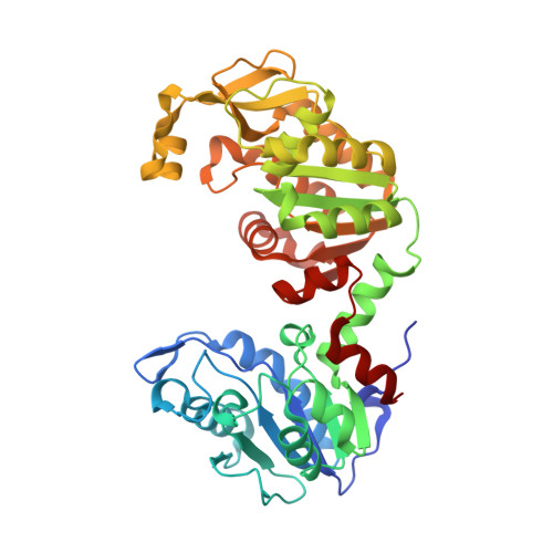 | |
UniProt | |||||
Find proteins for A0A0M3KL68 (Acinetobacter baumannii AB5075) Explore A0A0M3KL68 Go to UniProtKB: A0A0M3KL68 | |||||
Entity Groups | |||||
| Sequence Clusters | 30% Identity50% Identity70% Identity90% Identity95% Identity100% Identity | ||||
| UniProt Group | A0A0M3KL68 | ||||
Sequence AnnotationsExpand | |||||
Reference Sequence | |||||
| Length ( Å ) | Angle ( ˚ ) |
|---|---|
| a = 70.44 | α = 90 |
| b = 200.29 | β = 91.37 |
| c = 87.3 | γ = 90 |
| Software Name | Purpose |
|---|---|
| XSCALE | data scaling |
| PHENIX | refinement |
| PDB_EXTRACT | data extraction |
| XDS | data reduction |
| BALBES | phasing |
| Funding Organization | Location | Grant Number |
|---|---|---|
| National Institutes of Health/National Institute Of Allergy and Infectious Diseases (NIH/NIAID) | United States | -- |


