5BSY | pdb_00005bsy

Crystal structure of GII.10 P domain in complex with lemon juice


Experimental Data Snapshot

  • Method: X-RAY DIFFRACTION
  • Resolution: 1.60 Å
  • R-Value Free: 
    0.189 (Depositor), 0.196 (DCC) 
  • R-Value Work: 
    0.160 (Depositor), 0.170 (DCC) 
  • R-Value Observed: 
    0.162 (Depositor) 

Starting Model: experimental
View more details

wwPDB Validation 3D Report Full Report

Validation slider image for 5BSY

This is version 1.1 of the entry. See complete history

Literature

Treatment of norovirus particles with citrate.

Koromyslova, A.D.White, P.A.Hansman, G.S.

(2015) Virology 485: 199-204

  • DOI: https://doi.org/10.1016/j.virol.2015.07.009
  • Primary Citation Related Structures: 
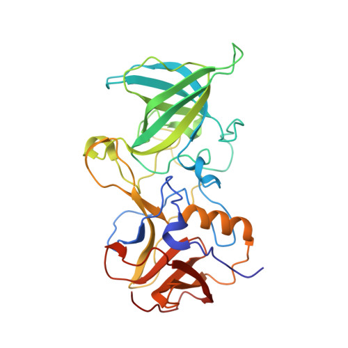
    5BSX, 5BSY

  • PubMed Abstract: 

    Human norovirus is a dominant cause of acute gastroenteritis around the world. Several norovirus disinfectants label citric acid as an active ingredient. In this study, we showed that norovirus virus-like particles (VLPs) treated with citrate buffer caused the particles to alter their morphology, including increased diameters associated with a new ring-like structure. We also found that epitopes on the protruding (P) domain on these particles were more readily accessible to antibodies after the citrate treatment. These results suggested that citrate had a direct effect on the norovirus particles. Using X-ray crystallography, we showed that the P domain bound citrate from lemon juice and a disinfectant containing citric acid. Importantly, citrate binds at the histo-blood group antigen binding pocket, which are attachment factors for norovirus infections. Taken together, these new findings suggested that it might be possible to treat/reduce norovirus infections with citrate, although further studies are needed.


  • Organizational Affiliation
    • Schaller Research Group at the University of Heidelberg and the DKFZ, Heidelberg 69120, Germany; Department of Infectious Diseases, Virology, University of Heidelberg, Heidelberg 69120, Germany.

Macromolecule Content 

  • Total Structure Weight: 69.57 kDa 
  • Atom Count: 5,548 
  • Modeled Residue Count: 624 
  • Deposited Residue Count: 630 
  • Unique protein chains: 1

Macromolecules

Find similar proteins by:|  3D Structure
Entity ID: 1
MoleculeChains  Sequence LengthOrganismDetailsImage
Capsid protein
A, B
315Norwalk virusMutation(s): 0 
UniProt
Find proteins for Q5F4T5 (Norwalk virus)
Explore Q5F4T5 
Go to UniProtKB:  Q5F4T5
Entity Groups
Sequence Clusters30% Identity50% Identity70% Identity90% Identity95% Identity100% Identity
UniProt GroupQ5F4T5
Sequence Annotations
Expand
Reference Sequence

Small Molecules

Ligands 3 Unique
IDChains Name / Formula / InChI Key2D Diagram3D Interactions
FLC

Query on FLC


Download:Ideal Coordinates CCD File
E [auth A]CITRATE ANION
C6 H5 O7
KRKNYBCHXYNGOX-UHFFFAOYSA-K
EDO

Query on EDO


Download:Ideal Coordinates CCD File
C [auth A],
D [auth A],
G [auth B],
H [auth B]
1,2-ETHANEDIOL
C2 H6 O2
LYCAIKOWRPUZTN-UHFFFAOYSA-N
NO3

Query on NO3


Download:Ideal Coordinates CCD File
F [auth A],
I [auth B]
NITRATE ION
N O3
NHNBFGGVMKEFGY-UHFFFAOYSA-N

Experimental Data & Validation

Experimental Data

  • Method: X-RAY DIFFRACTION
  • Resolution: 1.60 Å
  • R-Value Free:  0.189 (Depositor), 0.196 (DCC) 
  • R-Value Work:  0.160 (Depositor), 0.170 (DCC) 
  • R-Value Observed: 0.162 (Depositor) 
Space Group: P 1 21 1
Unit Cell:
Length ( Å )Angle ( ˚ )
a = 65.08α = 90
b = 79.75β = 100.98
c = 69.47γ = 90
Software Package:
Software NamePurpose
PHENIXrefinement
XSCALEdata scaling
PDB_EXTRACTdata extraction
PHASERphasing

Structure Validation

View Full Validation Report


Entry History 

Deposition Data

Revision History  (Full details and data files)

  • Version 1.0: 2015-09-09
    Type: Initial release
  • Version 1.1: 2024-01-10
    Changes: Data collection, Database references, Refinement description