5BSF | pdb_00005bsf

Crystal structure of Medicago truncatula (delta)1-Pyrroline-5-Carboxylate Reductase (MtP5CR) in complex with NAD+


Experimental Data Snapshot

  • Method: X-RAY DIFFRACTION
  • Resolution: 1.85 Å
  • R-Value Free: 
    0.177 (Depositor), 0.180 (DCC) 
  • R-Value Work: 
    0.155 (Depositor) 
  • R-Value Observed: 
    0.156 (Depositor) 

Starting Models: experimental
View more details

wwPDB Validation 3D Report Full Report

Validation slider image for 5BSF

Ligand Structure Quality Assessment 


This is version 1.2 of the entry. See complete history

Literature

The structure of Medicago truncatula delta (1)-pyrroline-5-carboxylate reductase provides new insights into regulation of proline biosynthesis in plants.

Ruszkowski, M.Nocek, B.Forlani, G.Dauter, Z.

(2015) Front Plant Sci 6: 869-869

  • DOI: https://doi.org/10.3389/fpls.2015.00869
  • Primary Citation Related Structures: 
    5BSE, 5BSF, 5BSG, 5BSH

  • PubMed Abstract: 

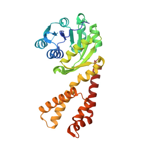
    The two pathways for proline biosynthesis in higher plants share the last step, the conversion of δ(1)-pyrroline-5-carboxylate (P5C) to L-proline, which is catalyzed by P5C reductase (P5CR, EC 1.5.1.2) with the use of NAD(P)H as a coenzyme. There is increasing amount of evidence to suggest a complex regulation of P5CR activity at the post-translational level, yet the molecular basis of these mechanisms is unknown. Here we report the three-dimensional structure of the P5CR enzyme from the model legume Medicago truncatula (Mt). The crystal structures of unliganded MtP5CR decamer, and its complexes with the products NAD(+), NADP(+), and L-proline were refined using x-ray diffraction data (at 1.7, 1.85, 1.95, and 2.1 Å resolution, respectively). Based on the presented structural data, the coenzyme preference for NADPH over NADH was explained, and NADPH is suggested to be the only coenzyme used by MtP5CR in vivo. Furthermore, the insensitivity of MtP5CR to feed-back inhibition by proline, revealed by enzymatic analysis, was correlated with structural features. Additionally, a mechanism for the modulation of enzyme activity by chloride anions is discussed, as well as the rationale for the possible development of effective enzyme inhibitors.


  • Organizational Affiliation
    • Synchrotron Radiation Research Section, Macromolecular Crystallography Laboratory, National Cancer Institute Argonne, IL, USA.

Macromolecule Content 

  • Total Structure Weight: 296.96 kDa 
  • Atom Count: 22,411 
  • Modeled Residue Count: 2,713 
  • Deposited Residue Count: 2,770 
  • Unique protein chains: 1

Macromolecules

Find similar proteins by:|  3D Structure
Entity ID: 1
MoleculeChains  Sequence LengthOrganismDetailsImage
Pyrroline-5-carboxylate reductase
A, B, C, D, E
A, B, C, D, E, F, G, H, I, J
277Medicago truncatulaMutation(s): 0 
Gene Names: MTR_7g090160
EC: 1.5.1.2
UniProt
Find proteins for G7KRM5 (Medicago truncatula)
Explore G7KRM5 
Go to UniProtKB:  G7KRM5
Entity Groups
Sequence Clusters30% Identity50% Identity70% Identity90% Identity95% Identity100% Identity
UniProt GroupG7KRM5
Sequence Annotations
Expand
Reference Sequence

Small Molecules

Ligands 3 Unique
IDChains Name / Formula / InChI Key2D Diagram3D Interactions
NAD

Query on NAD


Download:Ideal Coordinates CCD File
CA [auth G]
FA [auth H]
IA [auth I]
K [auth A]
LA [auth J]
CA [auth G],
FA [auth H],
IA [auth I],
K [auth A],
LA [auth J],
N [auth B],
Q [auth C],
T [auth D],
W [auth E],
Z [auth F]
NICOTINAMIDE-ADENINE-DINUCLEOTIDE
C21 H27 N7 O14 P2
BAWFJGJZGIEFAR-NNYOXOHSSA-N
MPO

Query on MPO


Download:Ideal Coordinates CCD File
AA [auth F]
DA [auth G]
GA [auth H]
JA [auth I]
L [auth A]
AA [auth F],
DA [auth G],
GA [auth H],
JA [auth I],
L [auth A],
MA [auth J],
O [auth B],
R [auth C],
U [auth D],
X [auth E]
3[N-MORPHOLINO]PROPANE SULFONIC ACID
C7 H15 N O4 S
DVLFYONBTKHTER-UHFFFAOYSA-N
CL

Query on CL


Download:Ideal Coordinates CCD File
BA [auth F]
EA [auth G]
HA [auth H]
KA [auth I]
M [auth A]
BA [auth F],
EA [auth G],
HA [auth H],
KA [auth I],
M [auth A],
NA [auth J],
P [auth B],
S [auth C],
V [auth D],
Y [auth E]
CHLORIDE ION
Cl
VEXZGXHMUGYJMC-UHFFFAOYSA-M

Experimental Data & Validation

Experimental Data

  • Method: X-RAY DIFFRACTION
  • Resolution: 1.85 Å
  • R-Value Free:  0.177 (Depositor), 0.180 (DCC) 
  • R-Value Work:  0.155 (Depositor) 
  • R-Value Observed: 0.156 (Depositor) 
Space Group: P 1
Unit Cell:
Length ( Å )Angle ( ˚ )
a = 87.54α = 67.5
b = 100.78β = 85.86
c = 101.61γ = 89.78
Software Package:
Software NamePurpose
XSCALEdata scaling
REFMACrefinement
PDB_EXTRACTdata extraction
XDSdata reduction

Structure Validation

View Full Validation Report


Ligand Structure Quality Assessment 


Entry History 

Deposition Data

Revision History  (Full details and data files)

  • Version 1.0: 2015-11-11
    Type: Initial release
  • Version 1.1: 2015-12-02
    Changes: Database references
  • Version 1.2: 2023-09-27
    Changes: Data collection, Database references, Derived calculations, Refinement description