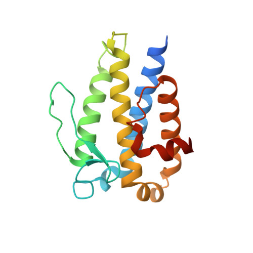
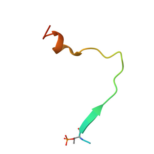
5BRM

Structural basis for Mob1-dependent activation of the core Mst-Lats kinase cascade in Hippo signaling


Experimental Data Snapshot

  • Method: X-RAY DIFFRACTION
  • Resolution: 2.65 Å
  • R-Value Free: 0.268 
  • R-Value Work: 0.244 
  • R-Value Observed: 0.245 

Starting Model: experimental
View more details

wwPDB Validation   3D Report Full Report


This is version 1.6 of the entry. See complete history


Literature

Structural basis for Mob1-dependent activation of the core Mst-Lats kinase cascade in Hippo signaling.

Ni, L.Zheng, Y.Hara, M.Pan, D.Luo, X.

(2015) Genes Dev 29: 1416-1431

  • DOI: https://doi.org/10.1101/gad.264929.115
  • Primary Citation of Related Structures:  
    5BRK, 5BRM

  • PubMed Abstract: 

    The Mst-Lats kinase cascade is central to the Hippo tumor-suppressive pathway that controls organ size and tissue homeostasis. The adaptor protein Mob1 promotes Lats activation by Mst, but the mechanism remains unknown. Here, we show that human Mob1 binds to autophosphorylated docking motifs in active Mst2. This binding enables Mob1 phosphorylation by Mst2. Phosphorylated Mob1 undergoes conformational activation and binds to Lats1. We determine the crystal structures of phospho-Mst2-Mob1 and phospho-Mob1-Lats1 complexes, revealing the structural basis of both phosphorylation-dependent binding events. Further biochemical and functional analyses demonstrate that Mob1 mediates Lats1 activation through dynamic scaffolding and allosteric mechanisms. Thus, Mob1 acts as a phosphorylation-regulated coupler of kinase activation by virtue of its ability to engage multiple ligands. We propose that stepwise, phosphorylation-triggered docking interactions of nonkinase elements enhance the specificity and robustness of kinase signaling cascades.


  • Organizational Affiliation

    Department of Pharmacology, University of Texas Southwestern Medical Center, Dallas, Texas 75390, USA;


Macromolecules
Find similar proteins by:  (by identity cutoff)  |  3D Structure
Entity ID: 1
MoleculeChains Sequence LengthOrganismDetailsImage
MOB kinase activator 1A
A, B, C, D, E
A, B, C, D, E, F
177Homo sapiensMutation(s): 0 
Gene Names: MOB1AC2orf6MOB4BMOBK1BMOBKL1B
UniProt & NIH Common Fund Data Resources
Find proteins for Q9H8S9 (Homo sapiens)
Explore Q9H8S9 
Go to UniProtKB:  Q9H8S9
PHAROS:  Q9H8S9
GTEx:  ENSG00000114978 
Entity Groups  
Sequence Clusters30% Identity50% Identity70% Identity90% Identity95% Identity100% Identity
UniProt GroupQ9H8S9
Sequence Annotations
Expand
  • Reference Sequence
Find similar proteins by:  (by identity cutoff)  |  3D Structure
Entity ID: 2
MoleculeChains Sequence LengthOrganismDetailsImage
Serine/threonine-protein kinase 3
G, H, I, J, K
G, H, I, J, K, L, M, N, O
31Homo sapiensMutation(s): 0 
EC: 2.7.11.1
UniProt & NIH Common Fund Data Resources
Find proteins for Q13188 (Homo sapiens)
Explore Q13188 
Go to UniProtKB:  Q13188
PHAROS:  Q13188
GTEx:  ENSG00000104375 
Entity Groups  
Sequence Clusters30% Identity50% Identity70% Identity90% Identity95% Identity100% Identity
UniProt GroupQ13188
Sequence Annotations
Expand
  • Reference Sequence
Experimental Data & Validation

Experimental Data

  • Method: X-RAY DIFFRACTION
  • Resolution: 2.65 Å
  • R-Value Free: 0.268 
  • R-Value Work: 0.244 
  • R-Value Observed: 0.245 
  • Space Group: P 31 2 1
Unit Cell:
Length ( Å )Angle ( ˚ )
a = 142.188α = 90
b = 142.188β = 90
c = 135.981γ = 120
Software Package:
Software NamePurpose
PHENIXrefinement
HKL-2000data scaling
PDB_EXTRACTdata extraction
PHENIXphasing

Structure Validation

View Full Validation Report


Entry History & Funding Information

Deposition Data

  • Released Date: 2015-07-08 
  • Deposition Author(s): Luo, X., Ni, L.

Funding OrganizationLocationGrant Number
National Institutes of Health/National Institute of General Medical Sciences (NIH/NIGMS)United StatesGM085004

Revision History  (Full details and data files)

  • Version 1.0: 2015-07-08
    Type: Initial release
  • Version 1.1: 2015-07-15
    Changes: Source and taxonomy
  • Version 1.2: 2015-07-22
    Changes: Database references
  • Version 1.3: 2017-09-20
    Changes: Author supporting evidence, Database references, Derived calculations, Source and taxonomy
  • Version 1.4: 2019-12-25
    Changes: Author supporting evidence
  • Version 1.5: 2023-09-27
    Changes: Data collection, Database references, Derived calculations, Refinement description
  • Version 1.6: 2024-11-20
    Changes: Structure summary